
Table of Content
- What is the physics behind Computed Radiography?
- Computed Radiography vs. Digital Radiography
- Computed Radiography as an NDT Technique
- Fundamentals of Computed Radiography
- Basic Principles of Computed Radiography (CR)
- Comparison to Conventional Radiography
- Components of a Typical Computed Radiography System
- Advantages of Computed Radiography over Conventional Film-Based Radiography
- Applications of Computed Radiography
- Image Interpretation and Defect Detection
- Steps Involved in Acquiring Computed Radiography Images
- Importance of Proper Technique Selection, Exposure, and Positioning
- Role of Image Quality Indicators (IQIs) and Their Interpretation
- Advantages of Computed Radiography in NDT Applications
- Limitations and Challenges of Computed Radiography Technology
- Mitigation and Overcoming Limitations
- Quality Assurance in Computed Radiography Imaging and Standards
- Role of Personnel Qualification and Training
- Maintenance and Care of Computed Radiography Equipment
- Common Issues and Troubleshooting Techniques
- Future Trends in Computed Radiography
- Future Evolution and Potential Impact on the Industry
- Computed Radiography in the Broader Landscape of NDT Techniques
- Final Words
Computed Radiography (CR) is an advanced imaging technique widely used in Non-destructive Testing (NDT) to detect internal defects and flaws in various materials and components.
It involves the use of imaging plates that capture radiation transmitted through the test specimen, producing digital images that can be processed and analyzed on a computer.
Computed Radiography has become an indispensable tool in many industries due to its ability to provide high-quality radiographic images without the need for traditional film-based methods.
What is the physics behind Computed Radiography?
Computed Radiography (CR) is an imaging technique used in medical radiography and Non-destructive Testing.
It involves capturing X-ray images using a phosphor plate that stores the X-ray energy temporarily.
When exposed to X-rays, the phosphor traps energy proportional to the radiation intensity.
Later, the plate is scanned with a laser beam, releasing the stored energy as visible light.
The emitted light is detected, converted into an electronic signal, and transformed into a digital image using computer algorithms.
Computed Radiography enables efficient image storage, retrieval, and manipulation, offering enhanced diagnostic capabilities while reducing patient exposure to radiation compared to traditional film-based radiography.
Computed Radiography vs. Digital Radiography
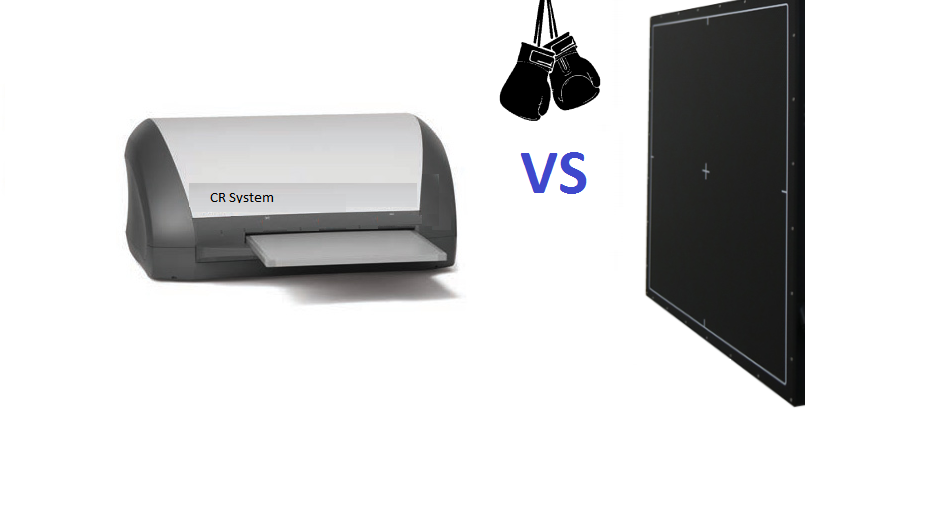
Computed Radiography (CR) and Digital Radiography (DR) are both modern imaging technologies used in radiology, offering significant advantages over traditional film-based Radiography.
However, they differ in their image capture and processing methods.
Computed Radiography utilizes a phosphor plate to capture X-ray images.
The plate temporarily stores the X-ray energy, which is later scanned with a laser beam to release the stored energy as visible light.
The emitted light is then converted into a digital image using computer algorithms.
Computed Radiography systems are an upgrade from analog film but require a separate reader for processing, which can lead to longer image acquisition times.
On the other hand, Digital Radiography directly captures X-ray images using electronic sensors.
These sensors, typically amorphous selenium or cesium iodide detectors, convert X-rays into electrical signals that are immediately processed and converted into digital images without the need for additional scanning.
Digital Radiography offers real-time image acquisition and eliminates the need for chemical processing, resulting in quicker image production and reduced patient exposure to radiation.
In summary, while both CR and DR provide digital images for efficient storage and manipulation, DR has the advantage of faster image acquisition and direct digital conversion, making it a more popular choice in modern radiology due to its improved workflow and reduced radiation dose to patients.
Computed Radiography as an NDT Technique
Non-destructive Testing (NDT) is a crucial set of techniques used to inspect and evaluate materials and components without causing damage to them.
Its significance lies in ensuring the integrity, safety, and quality of various industrial assets.
NDT is employed in industries such as aerospace, automotive, oil and gas, manufacturing, and construction.
Radiographic Testing is a fundamental NDT method that uses X-rays or gamma rays to examine the internal structure of objects.
It is particularly valuable for inspecting welds, castings, and other critical components, as it can reveal defects and discontinuities not visible to the naked eye.
Computed Radiography (CR) is an advanced NDT technique within radiographic testing.
It involves using a phosphor plate to capture X-ray images. This plate stores X-ray energy temporarily, which is later scanned and converted into a digital image using computer algorithms.
Computed Radiography offers numerous benefits, including efficient image storage, retrieval, and manipulation, as well as reduced radiation exposure for patients and technicians compared to traditional film-based radiography.
Fundamentals of Computed Radiography
Computed Radiography (CR) is an advanced Non-destructive Testing (NDT) technique used to capture X-ray images digitally.
It involves the use of a flexible imaging plate coated with a phosphor material that temporarily stores X-ray energy when exposed to radiation.
The stored energy forms a latent image on the plate, which can later be extracted and processed to produce a digital radiographic image.
Don't Miss Out, Radiographic Films: A Complete Guide to Understanding the Basics
Basic Principles of Computed Radiography (CR)
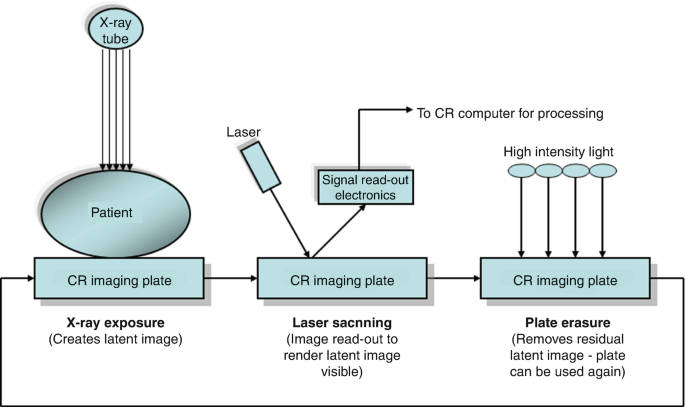
1. Exposure
The imaging plate is placed behind the object being tested, and X-rays are directed through the object onto the plate.
The X-rays penetrate the object, leaving varying levels of energy on the plate based on the object's internal structure and density.
2. Latent Image Formation
The phosphor in the imaging plate absorbs the X-ray energy and stores it as a latent image.
The distribution of energy corresponds to the object's internal features.
3. Scanning
After exposure, the imaging plate is removed from the X-ray environment and inserted into a Computed Radiography scanner.
A laser beam scans the plate, releasing the stored energy as visible light.
4. Light Conversion
The emitted light is detected by photomultiplier tubes or similar sensors, converting the light signals into electronic signals.
5. Digital Image Creation
The electronic signals are digitized and processed by a computer, creating a digital radiographic image that can be viewed, stored, and analyzed.
Comparison to Conventional Radiography
In conventional radiography, X-ray images are captured on film, which requires chemical processing to develop the images.
This process is time-consuming, and the images need physical storage.
In contrast, CR offers digital imaging, allowing for immediate image acquisition, easy storage, retrieval, and sharing.
Computed Radiography also reduces the need for repeat exposures due to under or overexposure, resulting in reduced radiation dose to patients and faster diagnosis.
Components of a Typical Computed Radiography System
1. Imaging Plate:
A flexible and reusable phosphor-coated plate that temporarily stores X-ray energy to form the latent image.
2. Scanner:
The device is used to read the imaging plate by scanning it with a laser beam and capturing the emitted light.
3. Reader:
The component detects the emitted light and converts it into electronic signals for further processing.
4. Computer System:
The computer and associated software digitize and process the electronic signals into a digital radiographic image.
Advantages of Computed Radiography over Conventional Film-Based Radiography
1. Digital Imaging:
CR provides digital radiographic images that can be instantly viewed, analyzed, and shared electronically, leading to faster and more efficient workflow.
2. Storage and Retrieval:
Digital images can be stored electronically, eliminating the need for physical film storage and making image retrieval easier and faster.
3. Image Manipulation:
Digital images allow for easy post-processing, enhancing image quality and aiding in the diagnostic process.
4. Reduced Radiation Exposure:
CR often requires lower X-ray exposure, reducing the radiation dose received by patients and operators.
5. Cost-Efficient:
Though initial setup costs may be higher, CR becomes cost-efficient in the long run due to reusable imaging plates and decreased chemical and film expenses.
Overall, Computed Radiography significantly improves the efficiency, safety, and quality of radiographic testing when compared to conventional film-based radiography.
Applications of Computed Radiography
Computed Radiography (CR) finds extensive applications in a wide range of industries, where Non-destructive Testing (NDT) is essential for ensuring the integrity, safety, and quality of critical components and structures.
Some of the industries benefiting from CR in NDT include:
1. Aerospace
- Applications: CR is used for inspecting aircraft components like turbine blades, engine parts, and composite materials to detect internal defects and ensure structural integrity.
- Advantages: CR's ability to reveal intricate internal details allows for accurate flaw detection, leading to improved reliability and safety of aircraft components.
2. Automotive
- Applications: CR is employed in automotive manufacturing to inspect welds, castings, and engine components to detect porosity, cracks, and other defects.
- Advantages: Faster inspection and detailed imaging enhance the quality control process, leading to better-performing and safer automotive parts.
3. Oil and Gas
- Applications: In the oil and gas industry, CR is used to inspect pipelines, pressure vessels, and welds in offshore structures to identify corrosion and defects.
- Advantages: CR's digital imaging capabilities allow for efficient inspection in remote locations, reducing inspection time and minimizing downtime.
4. Manufacturing
- Applications: CR is crucial for inspecting welds and components in various manufacturing sectors, including heavy machinery, electronics, and consumer goods.
- Advantages: Quick and accurate inspections enhance manufacturing efficiency, reduce production delays, and maintain product quality.
5. Power Generation
- Applications: CR is employed in power plants to inspect critical components, such as turbines and steam generators, to detect flaws and ensure safety.
- Advantages: CR's digital format allows for easy archiving and comparison of images over time, facilitating predictive maintenance and ensuring long-term plant reliability.
6. Construction
- Applications: CR is used for inspecting welds and concrete structures in buildings and bridges to assess the integrity and identify potential weaknesses.
- Advantages: CR's ability to detect hidden defects aids in preventing structural failures and contributes to safer and more durable constructions.
7. Railways
- Applications: CR is utilized to inspect rail tracks, locomotives, and railway components to detect cracks and defects.
- Advantages: Efficient and reliable inspections enhance railway safety and reduce the risk of accidents.
Overall, CR's adoption in various industries for NDT applications has revolutionized quality control and inspection processes.
The advantages of CR, including digital imaging, faster inspections, and improved defect detection, lead to increased safety, enhanced productivity, and cost savings for these industries.
By detecting flaws early and accurately, CR helps prevent catastrophic failures, extends the lifespan of critical components, and ensures that products and structures meet the highest quality standards.
Image Interpretation and Defect Detection
Image interpretation and flaw identification are essential procedures in the current digital era.
They entail deriving useful information from visual data and spotting flaws.
These technologies have numerous uses, ranging from manufacturing quality control to medical imaging.
Steps Involved in Acquiring Computed Radiography Images
Here are the steps that should be followed for acquiring CR Images.
1. Technique Selection:
Determine the appropriate CR technique based on the material, component, and inspection requirements.
Factors like material thickness, density, and expected defects influence the choice of X-ray energy, exposure time, and imaging plate sensitivity.
2. Exposure:
Position the imaging plate behind the object to be inspected and expose it to X-rays.
The X-rays penetrate the object and create a latent image on the imaging plate, capturing the internal features.
3. Imaging Plate Handling:
After exposure, carefully remove the imaging plate from the X-ray environment to avoid any unintentional radiation exposure.
4. Scanning:
Place the exposed imaging plate into the CR scanner.
A laser beam scans the imaging plate, releasing the stored X-ray energy as visible light.
5. Light Conversion:
Photomultiplier tubes or similar sensors detect the emitted light and convert it into electronic signals.
6. Image Digitization:
The electronic signals are digitized and processed by a computer to create a digital radiographic image.
The image can now be displayed and analyzed on a monitor.
Importance of Proper Technique Selection, Exposure, and Positioning
- Proper technique selection ensures that the image captures the necessary details and defects with appropriate image contrast and resolution.
- Accurate exposure prevents underexposure (which may lead to missed defects) or overexposure (which can obscure defects).
- Correct positioning ensures that the desired area of interest is adequately represented in the image, minimizing artifacts and enhancing defect detection.
Role of Image Quality Indicators (IQIs) and Their Interpretation
- IQIs are used to assess the image quality and performance of the CR system. They contain different thicknesses of materials, which, when visible in the image, provide information about the sensitivity and resolution of the system.
- The visibility of IQIs indicates whether the system is capable of detecting specific sizes of defects. If an IQI's details are not visible in the image, it may suggest the need for adjustments in the technique or positioning to improve image quality.
Examination Process, Including Image Enhancement and Manipulation
- After acquiring the CR image, inspectors review it for defects, discontinuities, or irregularities. Image enhancement techniques may be applied to improve visibility, such as adjusting contrast, brightness, or gamma correction.
- Image manipulation can involve zooming, panning, or filtering to focus on specific areas of interest for closer inspection. Annotations and measurements may be added to highlight and quantify defects.
- Comparison with standards or previous images allows inspectors to identify changes over time or deviations from acceptable criteria.
The examination process should involve qualified personnel with expertise in interpreting NDT images.
Proper technique selection, exposure, and positioning, along with the use of IQIs, ensure reliable and accurate results, aiding in the detection and assessment of defects.
CR's digital format facilitates image enhancement and manipulation, providing a comprehensive evaluation of inspected components and supporting informed decision-making for maintenance, repairs, and quality control in various industries.
Advantages of Computed Radiography in NDT Applications
1. Digital Imaging:
CR offers digital radiographic images, enabling immediate viewing, analysis, and storage.
This leads to faster NDT Inspection Processes and efficient data management.
2. Image Manipulation:
Digital images allow for easy enhancement, zooming, and measurement, improving defect visualization and analysis.
3. Reduced Radiation Exposure:
CR typically requires lower X-ray exposure compared to conventional Film-based Radiography, reducing radiation dose to both patients and operators.
4. Cost-Efficiency:
While CR setup costs may be higher, the reusability of imaging plates and reduced chemical and film expenses make it cost-effective in the long run.
5. Enhanced Defect Detection:
CR's high sensitivity and resolution enable the detection of smaller or hidden defects, leading to improved reliability and safety in inspected components.
Limitations and Challenges of Computed Radiography Technology
1. Initial Investment
Setting up a CR system can involve higher initial costs for the equipment and training.
2. Plate Handling Sensitivity
Imaging plates are sensitive to light, heat, and physical damage, requiring careful handling and storage.
3. Scanning Time
The scanning process can be time-consuming, impacting inspection throughput, especially when dealing with a large number of images.
4. Resolution Limitation
The resolution of CR images might not be as high as some other Digital Radiographic Methods, potentially affecting fine defect detection.

Mitigation and Overcoming Limitations
1. Proper Training
Ensure technicians are well-trained in CR operation, imaging plate handling, and accurate image interpretation to maximize efficiency and accuracy.
2. Optimize Workflow
Plan and optimize the scanning process to minimize downtime and maximize inspection throughput.
3. Quality Assurance
Implement regular quality checks, calibration, and maintenance of CR equipment to ensure reliable and consistent results.
4. Consider Image Stitching
In cases where resolution limitations are critical, consider stitching multiple images together to create a larger, more detailed image.
5. Supplement with Other Techniques
Depending on specific requirements, consider supplementing CR with complementary NDT methods, such as Ultrasonic Testing or digital radiography, to enhance defect detection capabilities.
6. Improve Plate Handling
Implement proper storage conditions and handling procedures for imaging plates to extend their lifespan and reduce the risk of damage.
By addressing these limitations and challenges through proper training, workflow optimization, and equipment maintenance, CR can provide significant benefits in NDT Applications.
The advantages of digital imaging, improved defect detection, reduced radiation exposure, and cost-efficiency make CR a valuable tool for ensuring the integrity, safety, and quality of components in various industries.

Quality Assurance in Computed Radiography Imaging and Standards
Quality assurance (QA) is critical in CR imaging to ensure reliable and accurate results in Non-destructive Testing (NDT) applications.
QA measures help maintain the performance and integrity of the CR system, minimize errors, and enhance the overall efficiency of the inspection process.
By implementing effective quality assurance practices, organizations can confidently assess the quality and safety of inspected components, contributing to better decision-making and risk mitigation.
Relevant International Standards and Guidelines for CR in NDT
ISO 17636:
This standard provides guidelines for the radiographic examination of metallic materials using X- and gamma-ray techniques.
It covers image quality indicators (IQIs), sensitivity, exposure techniques, and acceptance criteria.
ASTM E2446:
This specification outlines the minimum requirements for acquiring, processing and interpreting CR images for NDT purposes.
It also addresses image quality and image quality indicator selection.
EN 14784:
This European Standard specifies general principles for industrial computed radiography systems for Non-destructive Testing.
ASME BPVC Section V:
The American Society of Mechanical Engineers (ASME) Boiler and Pressure Vessel Code, Section V, includes guidelines for radiographic examination, covering CR and other Radiographic Methods.
Role of Personnel Qualification and Training
1. Qualified Personnel
Properly trained and qualified personnel are essential for conducting CR inspections accurately and efficiently.
Qualified inspectors possess the necessary knowledge and skills to operate CR equipment, interpret images, and follow proper NDT Inspection Procedures.
2. Training Programs
Organizations should invest in comprehensive training programs for CR personnel.
Training should cover CR system operation, calibration, image acquisition, interpretation, and quality control procedures.
3. Certification
Obtaining certification from recognized NDT Certification bodies helps validate the competence of CR personnel and ensures compliance with international standards.
4. Continuous Learning
The field of NDT and CR technology is constantly evolving.
Personnel should engage in ongoing education and participate in professional development activities to stay updated with the latest advancements and best practices.
5. Quality Management System
Implementing a quality management system helps maintain consistency and standardization in CR imaging practices.
Regular audits and reviews ensure adherence to established procedures and standards.
Maintenance and Care of Computed Radiography Equipment

* Guidelines for Proper Maintenance and Care of CR Equipment
Maintaining and caring for CR equipment is essential to ensure its longevity, accuracy, and optimal performance.
Follow these guidelines to keep your CR equipment in top condition:
1. Regular Inspection
Conduct regular inspections to identify any signs of wear, damage, or malfunction. Address issues promptly to prevent further damage.
2. Keep a Clean Environment
Maintain a clean and dust-free environment around the CR equipment to prevent particles from interfering with imaging plate performance.
3. Proper Storage
When not in use, store imaging plates in their protective envelopes or cassettes to prevent physical damage and light exposure.
4. Calibration and Service Schedule
Follow the manufacturer's recommended calibration and service schedule to maintain accurate imaging and prevent issues.
5. Operator Training
Ensure that all personnel operating the CR equipment receive proper training to minimize operator-induced errors and mishandling.
* Routine Checks and Cleaning Procedures
1. Imaging Plate Inspection
Regularly check imaging plates for scratches, cracks, or other physical damage. Damaged plates should be replaced promptly.
2. Cassette Integrity
Inspect cassettes for any damage or warping that might affect proper sealing and light tightness.
3. Cleaning CR Scanner
Keep the CR Scanner clean and free from debris. Use a soft, lint-free cloth to wipe surfaces gently, avoiding any harsh cleaning agents.
4. Cassette Cleaning
Clean cassettes with a soft cloth and mild cleaning solution as needed, ensuring they are thoroughly dry before use.

5. Imaging Plate Handling
Handle imaging plates with care and avoid touching the sensitive surface to prevent smudging and contamination.
Calibration Practices
1. Regular Calibration
Follow the manufacturer's recommended calibration schedule to maintain accurate image acquisition.
2. Reference Images
Maintain a library of reference images of known defects and indications for consistent calibration and sensitivity checks.
3. IQI Calibration
Periodically verify the imaging system's sensitivity using Image Quality Indicators (IQIs) of known thicknesses and sizes.

Common Issues and Troubleshooting Techniques
1. Image Artifacts
If artifacts appear in CR images, check for dust or debris on the imaging plate or scanner.
Cleaning and proper handling can resolve this issue.
2. Inconsistent Image Quality
Ensure consistent exposure to NDT Techniques, positioning, and image processing to maintain uniform image quality.
3. Scanner Malfunction
If the CR scanner experiences issues, consult the manufacturer's troubleshooting guide or contact their technical support.
4. Software Errors
Regularly update CR software and address any software-related errors promptly.
5. Plate Errors
If imaging plates show abnormalities, inspect for damage, proper handling, and storage.
By following these guidelines for maintenance, cleaning, calibration, and troubleshooting, you can optimize the performance and longevity of your CR equipment, ensuring reliable and accurate results in Non-destructive Testing Applications.
Future Trends in Computed Radiography
Emerging Technologies and Advancements in CR for NDT

1. Enhanced Resolution and Sensitivity
Advances in CR imaging technology are expected to improve resolution and sensitivity, enabling the detection of smaller defects and finer details in inspected components.
2. Faster Scanning Speed
Ongoing research aims to reduce the scanning time for CR imaging, making inspections more efficient and enabling higher throughput.
3. Automated Image Analysis
AI-powered image analysis algorithms are being developed to assist NDT Inspectors in defect detection and interpretation, reducing human error and speeding up the Inspection Process.
4. Wireless Connectivity
Integration of wireless technology into CR systems may eliminate the need for physical connections and allow for remote operation and data transfer.
5. Hybrid Imaging Techniques
Combining CR with other NDT Methods, such as computed tomography (CT), could provide more comprehensive inspection capabilities and 3D visualization of internal structures.
Future Evolution and Potential Impact on the Industry
The future of CR in NDT is likely to be shaped by rapid technological advancements, driving the following potential impacts on the industry:
1. Increased Efficiency
Faster scanning, automated analysis, and improved imaging quality will lead to faster and more accurate NDT Inspections, reducing downtime and improving productivity in various industries.
2. Expanded Applications
With enhanced resolution and sensitivity, CR will be able to address more critical applications and materials, including thin-walled structures and complex geometries.

3. Cost-Effectiveness
Advancements in CR technology may lead to more affordable NDT Equipment and reduced operational costs, making it accessible to a wider range of industries.
4. Greater Safety and Reliability
Improved defect detection and characterization will enhance safety and reliability in critical components, such as those used in the Aerospace Industry, transportation, and energy sectors.
Computed Radiography in the Broader Landscape of NDT Techniques
Computed Radiography is a versatile and complementary NDT Technique that coexists with other methods, providing its unique advantages:
1. Digital Imaging
CR offers digital images that facilitate efficient storage, retrieval, and analysis, setting it apart from conventional Film-based Radiography.
2. Radiographic Inspection
CR is well-suited for inspecting welds, castings, and composite materials, complementing other Radiographic Methods like X-ray and gamma-ray radiography.
3. Advanced Non-destructive Testing
CR's evolving technology and emerging applications contribute to the broader landscape of Advanced NDT Techniques, offering enhanced capabilities for defect detection and characterization.
As Computed Radiography continues to advance, its integration with other NDT Methods, automation, and increased accessibility will play a vital role in maintaining the integrity and safety of critical assets across various industries
As a key component of the NDT toolbox, Computed Radiography will continue to evolve and impact the industry by providing reliable and efficient solutions for inspecting and ensuring the quality of components and structures.

Final Words
Computed Radiography (CR) has emerged as a powerful and advanced Non-destructive Testing (NDT) Technique, revolutionizing the field of radiographic inspection.
Its digital imaging capabilities offer numerous advantages over conventional film-based Radiography, such as faster image acquisition, efficient data management, and reduced radiation exposure.
With the rapid pace of technological advancements, embracing the potential of CR and staying abreast of the latest developments will empower NDT Professionals to overcome challenges, improve inspection processes, and make informed decisions in critical sectors where safety and quality are paramount.
By embracing CR's digital imaging capabilities and combining them with expert analysis and industry best practices, we can continue to push the boundaries of Non-destructive Testing, ensuring a safer and more reliable future for diverse industries and applications.
References
1. Heditech


