Table of Content
- Basics of Radiographic Films
- Types of Radiographic Films
- Factors Considered When Selecting Radiographic Films
- Processing of Radiographic Films
- Packaging and Handling of Radiographic Films
- Applications of Radiographic Films
- Key Takeaways
Radiographic films serve as indispensable tools in various fields, particularly medicine, and industry, for capturing images produced by the passage of X-rays or gamma rays through objects. These films play a vital role in medical imaging, allowing healthcare professionals to diagnose and treat various medical conditions by visualising internal structures.
Basics of Radiographic Films
Radiographic films are sensitive to X-rays and gamma rays, producing images that can be used for diagnostic purposes in medicine and industry. These films are typically composed of a base material coated with emulsion layers containing silver halide crystals.
Composition and Structure
Radiographic films usually have a base layer made of polyester or cellulose and are covered with an emulsion that has silver halide crystals mixed in with gelatin. Upon exposure to radiation, these crystals undergo chemical changes, forming latent images.
Sensitivity to Radiation
The sensitivity of radiographic films varies based on factors such as emulsion thickness and grain size. Higher sensitivity films require shorter exposure times, facilitating rapid imaging processes.
What are the Types of Radiographic Films?
Radiographic films can be categorised based on various characteristics:
1. Conventional Films
Conventional radiographic films utilise analogue technology and require chemical processing for image development. Despite the advent of digital radiography, conventional films remain relevant due to their reliability and affordability.
2. Digital Radiography
Digital radiography involves electronic detectors capturing and displaying X-ray images directly onto a computer screen. Digital Radiography technology offers immediate image availability, enhanced manipulation capabilities, and improved storage options.
3. Screen-Film Radiography
Screen-film radiography involves using intensifying screens in conjunction with radiographic films to enhance image quality. These screens amplify the effect of X-rays, resulting in shorter exposure times and improved contrast.
4. Direct Radiography
Direct radiography eliminates the need for film by capturing X-ray images directly onto digital sensors. This method offers high image resolution, rapid image acquisition, and reduced radiation exposure for patients.
5. Computed Radiography (CR)
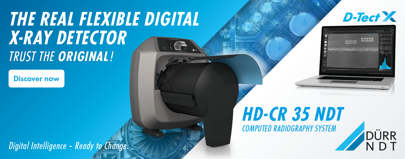
Computed radiography uses photostimulable phosphor plates to capture X-ray images, which are then processed digitally. Computed Radiography Systems provide flexibility in image manipulation and storage, making them suitable for various imaging applications.
6. Fluoroscopic Imaging
Fluoroscopic imaging involves real-time X-ray imaging of moving structures, such as the digestive system or blood vessels. Specialised fluoroscopic films are used to capture dynamic images during procedures like Angiography or barium studies.
7. Mammographic Imaging
Mammographic films are used for breast imaging to detect abnormalities such as tumours or cysts. These films are designed to provide high contrast and spatial resolution for accurate breast cancer screening.
8. Industrial Radiography
Industrial radiographic films are used for non-destructive testing in industries such as aerospace, automotive, and manufacturing. These films are designed to detect flaws and defects in materials and welds for quality control purposes.
These diverse types of radiographic films cater to various imaging needs across different fields, providing valuable insights into internal structures and abnormalities.
What are the factors considered when selecting Radiographic Films?
Different components have different requirements regarding their reaction to different ionizing radiation, different intensities of the resulting image, and varied reactions to the power applied. The factors that determine the quality and intensity of the resulting radiographic image are as follows:
- The physical characteristics and dimensions of the workpiece or test subject undergoing non-destructive inspection under radiographic techniques are of utmost concern when determining the best results.
- The ionizing rays impinged on the material (X-rays or gamma rays) also play a key role in ensuring optimal radiographic test results.
- Priority of the operator in terms of the quality of results versus the time constraint in obtaining them also plays an important role in the final result and selection of radiology films.
Processing of Radiographic Films
Further processing in the Radiographic Process is conducted after the change in the state of the materials suspended within the emulsion. To study this latent image, which is invisible to the human eye, the film is developed using the following three techniques i.e. Manual Method, Semi-Automatic Method, Automatic Method
The processing of films aids in making the particles suspended within the emulsion produce an image of higher intensity. The process consists of the following steps:
1. Developer
A developing agent is used under controlled temperature for a dwell period to convert the silver halide crystals to darker metallic silver for increased visibility. This image formed by metallic silver is called the manifest image. It should be ensured that the developer does not sit for longer than 12 minutes (or max time as specified for the developer in that environment), as it can convert the crystals unexposed to radiation to metallic silver as well.
2. Bath
This step processes the film in a water bath that slows off the excess developer. This, in turn, stops the previous process at a precise time sufficient for highlighting only the radiation-exposed particles to obtain accurate NDE Inspection results. This process only lasts around 15 seconds.
3. Fixing Process
This process involves another bath for approximately 5 minutes. This bath dissolves the unexposed silver halide crystals and leaves only the metallic silver. This step ensures the storage ability of the image obtained.
4. Wash
In this step, the film is washed with water to remove any chemicals used during its processing.
5. Preparation for viewing
To view the results, the film is either dried for ease of observation or is wet to swell the emulsion, depending on the material and other vital factors involved. Care should be taken to ensure uniformity of operation and precise control of environmental and testing factors involved in the process. The operator’s skill and thorough NDT Training ensure high-quality results.
Explore the Process of Radiographic Films Here!
Packaging and Handling of Radiographic Films
Radiographic films are commercially available in multiple forms. Selection of the right product is of utmost importance as it can affect how the resultant images are produced. The purchase of films in packaging most appropriate for the operator's intended method of use and handling style can make or break this NDE Process. In-depth knowledge of the intricacies of the process and thorough NDT Training can help an operator or organization choose between individual sheets available in a box or singularly packed sheets of radiographic films.
The individually packed sheets are packed in envelopes tightly protected from light. A mechanism like a ‘rip-strip’ is provided for easy removal from its packaging during usage to ensure no contamination and efficient use. This is incredibly easy to use and ensures easy access to the film in a darkroom. More commonly available film sheets are packed together in a box.
Each individual sheet requires careful handling and protection from light; hence it is housed in a cassette which is a film holder that protects the radiographic film from unwanted light exposure. For complex radiographic testing, wherein materials of cylindrical geometry etc., are to be analyzed, rolls of radiographic films are also available. This gives the operator the freedom to adjust the film length used. This can be economically viable in an industry wherein varied test subjects are observed under Radiography Testing.
Applications of Radiographic Films
Radiographic films play a critical role in industrial settings, particularly in Non-destructive Testing, where they are used to inspect materials and components for defects without causing damage. Here are the primary applications of radiographic films in industrial and NDT contexts:
1. Weld Inspection
Radiographic films are widely used to inspect welds in various industries, including construction, automotive, and aerospace. They help detect defects such as cracks, porosity, lack of fusion, and incomplete penetration in Welded Joints, ensuring structural integrity and quality control.
2. Casting and Forging Inspection
In manufacturing processes involving casting and forging, radiographic films are employed to examine the internal structure and integrity of components. They help identify defects such as shrinkage cavities, gas porosity, inclusions, and cracks, ensuring the quality of castings and forgings.
3. Pipeline Integrity Assessment
Radiographic films are used in the oil and gas industry to assess the Integrity of Pipelines and piping systems. They enable inspectors to detect corrosion, weld defects, and other anomalies that could compromise pipeline safety and reliability, helping prevent leaks and failures.
4. Pressure Vessel Testing
In industries such as petrochemicals, power generation, and manufacturing, radiographic films are used to inspect Pressure Vessels for Defects and weaknesses. They help ensure compliance with regulatory standards and prevent catastrophic failures that could result in safety hazards and environmental damage.
5. Aircraft Component Inspection
In the Aerospace Industry, radiographic films are employed to inspect critical aircraft components, including engine parts, landing gear, and structural elements. They help detect defects such as fatigue cracks, material degradation, and manufacturing flaws, ensuring airworthiness and safety.
6. Automotive Manufacturing
Radiographic films play a crucial role in automotive manufacturing, where they are used to inspect components such as engine blocks, chassis, and suspension systems. They help identify defects in castings, forgings, and welds, ensuring the quality and reliability of automotive vehicles.
7. Quality Control in Metalworking
In metalworking industries such as steel production and machining, radiographic films are employed for quality control purposes. They help identify internal defects and inconsistencies in metal components, ensuring they meet specified standards for strength, durability, and performance.
Radiography films can be delicate and require careful and trained handling. Any error on the operator’s part in the handling of the film can cause damage to the film and affect the results of the NDT Testing Process. Uniform pressure should be applied, and strain, friction, and scratches to the film should be avoided at all costs. The borders should handle the film to avoid surface contamination and scratches, which can affect the results irrespective of the technique or voltage used. Individually packed films can help avoid these issues.
Radiography is a boon to technology and can help detect defects deep within a subject. This penetrating nature can, however, be hazardous to human tissues and physical well-being. Operators and personnel should be educated about the hazards of being around these types of machinery and equipped with proper protective gear. Organizations that peruse such NDT Processes should be aware of the hazards that often come with such advanced technology and take an incentive to ensure the protection of human life and the environment while utilizing this technology to its best.
Key Takeaways
- Radiographic films continue to be indispensable tools in medical and industrial imaging, providing valuable insights into internal structures and anomalies.
- Despite advancements in digital imaging, conventional films remain relevant due to their reliability, affordability, and widespread availability.
- As technology evolves, radiographic imaging is expected to further enhance diagnostic capabilities, driving improvements in patient care and outcomes.
References
1. TWI Global
2. Pocket Dentistry
3. NDE-Ed.org
4. World of NDT
.png)


