
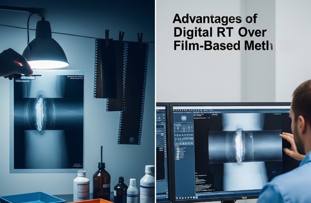
Digital radiography has changed the way industrial radiographic testing is performed. Direct image acquisition eliminates film processing, makes results available immediately after exposure and gives operators greater control over imaging parameters and subsequent analysis.
But faster acquisition is only one step toward a truly significant productivity boost. Once a radiograph has been obtained, one of the most demanding stages begins: interpretation. A specialist must examine the weld for relevant indications, classify and measure findings, determine their location and document the results. As working volumes grow, this creates a practical bottleneck. Throughput remains closely tied to the availability and experience of qualified personnel, while repetitive analytical work can contribute to variability between evaluations.
Assisted Defect Recognition (ADR) introduces automation at precisely this stage.
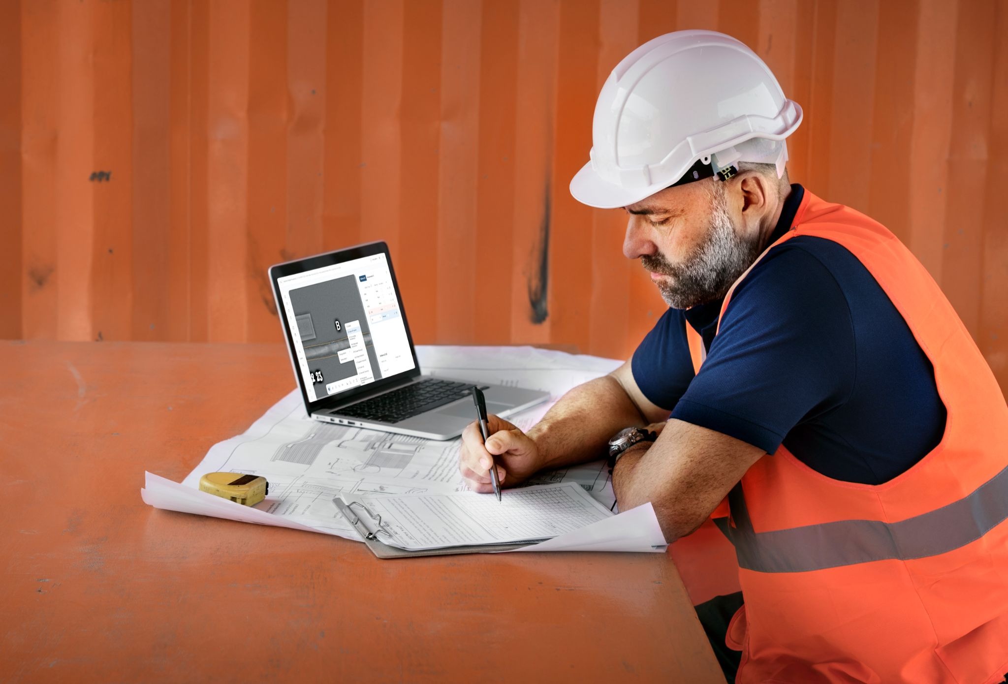
Developed by DR SYSTEMS specifically for digital radiographic inspection of welded joints, iMeva AI uses Computer Vision and Deep Learning to support the evaluation process from initial image analysis to final report generation. The objective is not to remove the qualified specialist from the process, but to automate routine operations and provide structured results for expert review.
From radiograph to evaluation
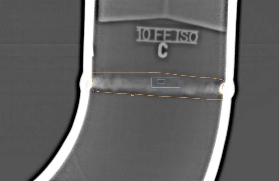
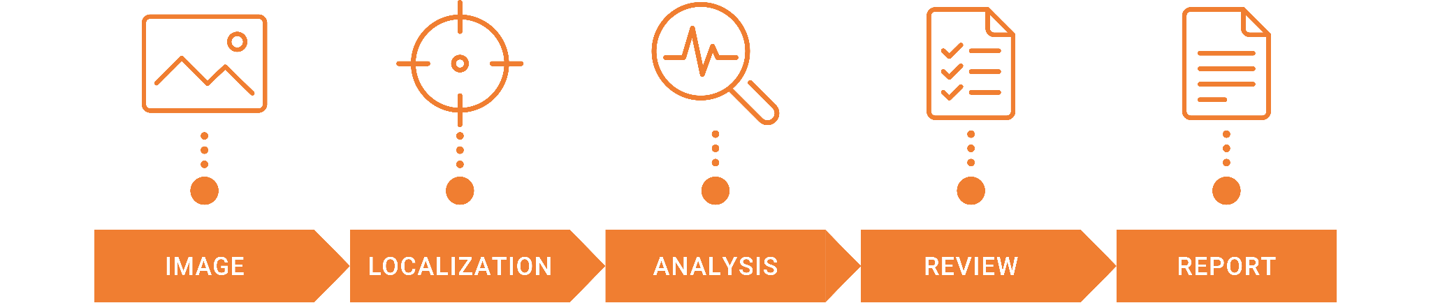
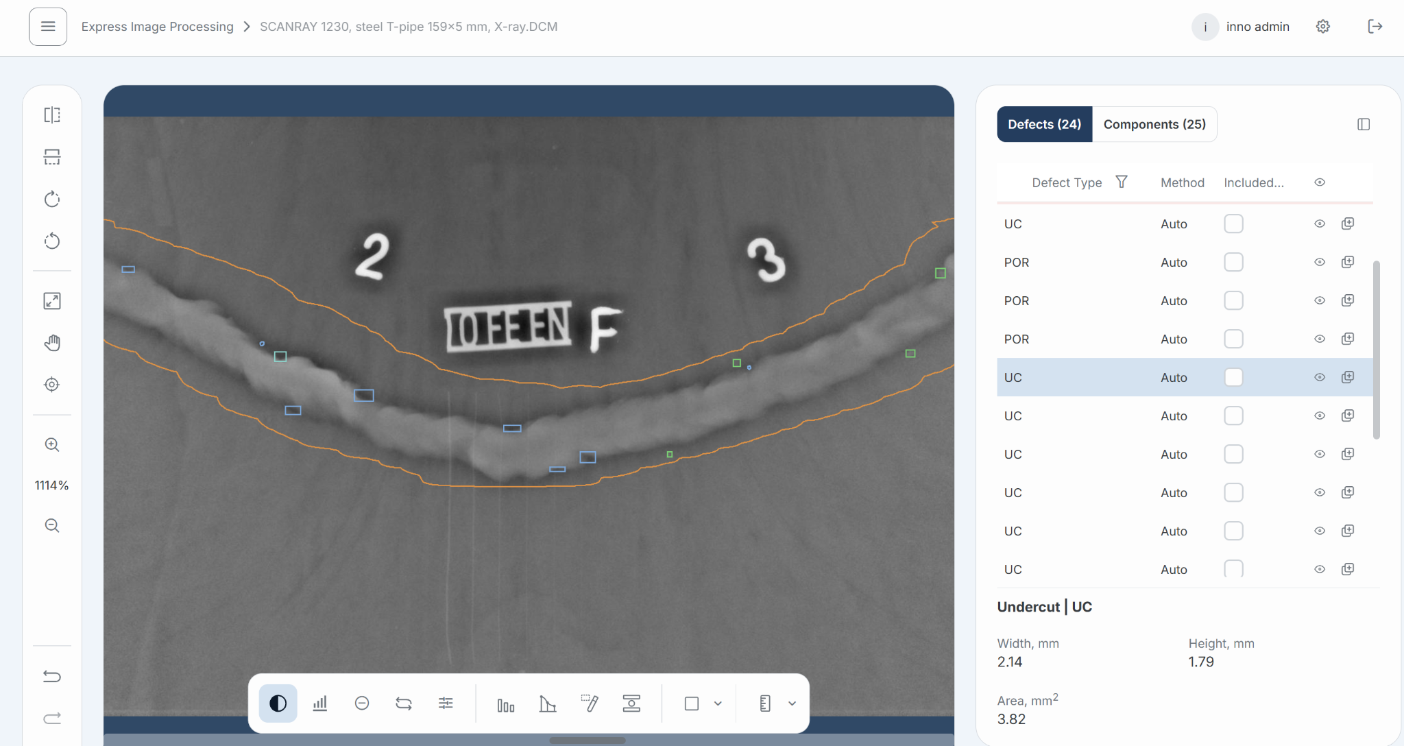
iMeva AI processes the image as a sequence of interconnected steps. DCM data and associated metadata are first validated and correlated. The software then identifies the weld contour and measuring belt, establishing the area in which subsequent analysis will be performed.
Then, the system searches for indications and determines their type, dimensions and coordinates. The findings are presented to the operator through an interface that provides filters, measurement and visualization tools for closer validation. Rather than producing a fixed AI verdict, the system creates an editable evaluation that the specialist can examine, correct where necessary and approve. This distinction is fundamental to the role of ADR. Automation handles a substantial part of the repetitive image-processing work, while responsibility for the final assessment remains with qualified personnel.
After confirmation, the system can generate a well-structured testing report with all flaw findings and the Approved/ Reject conclusion.
Recognizing the indications that matter
iMeva AI currently supports 15 defect categories used in welded-joint evaluation. These include common indications such as porosity and cluster porosity, undercut, incomplete fusion, inadequate penetration, cracks, slag indications and tungsten inclusions, alongside other discontinuity types.
For each detected indication, the software determines its position and physical dimensions and assigns a classification. The evaluation is performed in accordance with ASME B31.3 using the selected acceptance criteria.
The purpose is not simply to highlight suspicious areas on a radiograph. Classification, measurement and localization are performed within the same workflow, allowing the information generated during analysis to be carried forward into the inspection record.
What is behind the AI?
For any AI-based inspection technology, performance depends heavily on the data used to develop and validate the model. A large dataset alone is not sufficient: its relevance, annotation quality and verification are equally important.
iMeva AI has been trained on more than 380,000 labeled radiographic images, including over 120,000 defect samples representing a wide range of welded-joint indications.
The annotations follow a unified approach based on relevant industry codes and regulatory requirements. They are subjected to a three-stage verification process involving qualified operators, experienced Level III DR specialists and independent experts. This is intended to reduce inconsistencies in labeling and provide a reliable reference for model training.
Training and validation data are separated to provide an independent assessment of system performance. The qualification process involves thousands of radiographs, with results reviewed and cross-validated by NDT experts. Following each model retraining cycle, particular attention is given to validation performance before the updated model is introduced.
The dataset continues to expand as additional testing material becomes available, providing a basis for further refinement of detection and classification performance.
From automation to measurable results
The practical value of ADR becomes clearer when the complete evaluation cycle is considered.
In the performance data collected over the years in many countries, conventional manual evaluation typically takes approximately 15-20 minutes per joint. Automated processing with the software reduces the average evaluation time to around 2-3 minutes.
The resulting difference becomes particularly significant in high-volume projects. Compared with approximately 30-50 joints processed per shift through conventional evaluation, workflows supported by iMeva AI can reach 150-200 inspection reports per day. Report generation, which can otherwise extend the process considerably, is performed automatically once the results have been reviewed.
The system has demonstrated defect classification accuracy of up to 96% and detection of indications starting from 0.1 mm under applicable inspection conditions.
These improvements do not come from accelerating one isolated operation. Time is saved across several stages that would otherwise require long manual input.
Performance across different inspection conditions
Radiographic inspection is rarely performed under a single standardized field setup. Pipe diameter, wall thickness, radiation source, exposure technique and image quality can vary considerably between applications.
To illustrate the software in practice, two cases with contrasting testing parameters are presented below.
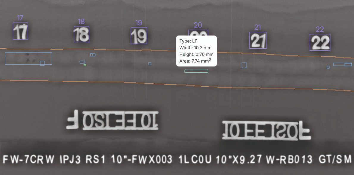
In one case, a 10-inch steel pipe with a wall thickness of 9.27 mm was inspected using the SCANRAY 1025F DR system, Comet EVO 200D X-ray source and the DWSI technique. Image quality of W14 (0.160 mm) was achieved. iMeva AI automatically recognized the weld and measuring belt and detected multiple indications, including lack of fusion, slag inclusions and several types of porosity, with measured dimensions ranging from 1.1 × 1.1 mm to 2.2 × 2.2 mm.
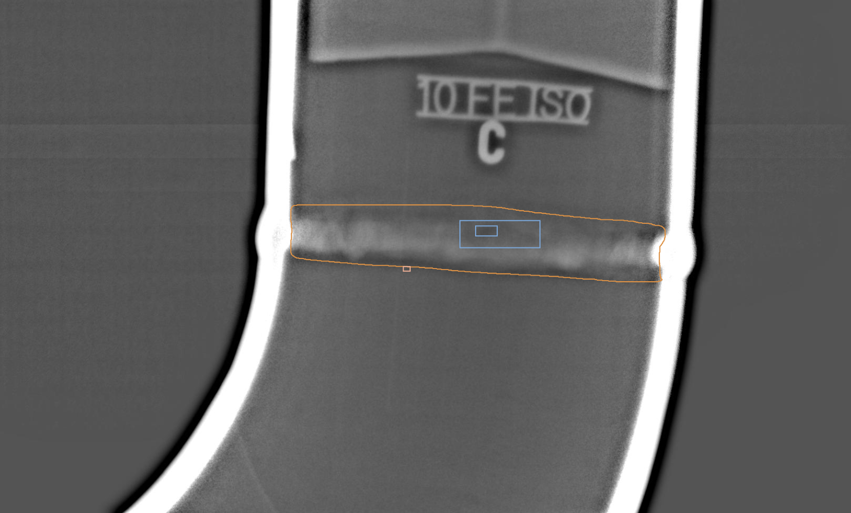
A second case involved a 3-inch pipe with a wall thickness of 5.49 mm, inspected with the SCANRAY 1230 DR system, an Ir-192 source and the DWDI superimposed technique. At an achieved image quality of W13 (0.200 mm), the software identified and measured undercut and internal concavity.
iMeva AI follows the approach that a truly universal industrial ADR solution cannot be tied to one idealized inspection setup; it must remain effective across the varied inputs encountered in routine inspection work.
Turning analysis into a structured inspection record
Finding an indication is not the end of the inspection process. Results still need to be documented in a form that connects image evaluation with the inspected object and acquisition conditions.
iMeva AI automatically generates a structured report containing weld identification, object and testing parameters, imaging conditions, as well as detected indications with their classifications and physical locations. Because the findings remain editable throughout the review stage, the report reflects the specialist's decision rather than an unverified AI output.
For larger organizations, this structured approach also creates a basis for centralized statistics, archives and traceability of user actions, rather than leaving evaluation data distributed across individual workstations and separate documents.
From a single workstation to enterprise deployment
The way ADR is introduced into an inspection organization depends on the scale and structure of its operations.
For standalone use, the iMeva AI Box Solution provides automated image evaluation and reporting on a local workstation without requiring complex additional infrastructure. It is intended for teams that need a ready-to-use environment for daily inspection work.
The Enterprise Solution uses a server-based architecture for organizations managing multiple users, projects or locations. It adds centralized data management, role-based access, synchronization, task management and options for integration with external systems.
In both configurations, sensitive testing information will always remain within the customer's own infrastructure rather than being transferred to external AI services. This is particularly relevant where radiographic data are subject to project-specific confidentiality or security requirements.
Bringing progress into practice
Digital radiography has already transformed how inspection data are acquired, accessed and managed. The next opportunity lies in making better use of the information those images contain.
iMeva AI by DR SYSTEMS takes this process further by bringing AI-assisted evaluation into everyday radiographic inspection. Instead of treating artificial intelligence as a future capability, it applies it to a working environment where image analysis and reporting can become faster, more consistent and easier to scale.
This marks another step in the broader digitalization of NDT. As inspection volumes grow and industrial processes become increasingly data-driven, progress will depend not only on acquiring high-quality images efficiently, but also on turning them into useful results with less manual effort.
By bringing these capabilities into practical use today, iMeva AI advances the way radiographic evaluation is performed and moves the industry closer to a more connected, automated and efficient approach to non-destructive testing.

There are no comments yet!