Table Of Contents
- Introduction
- The Basic Principles of Computed Radiography
- Function of Computed Radiography (CR) in Radiography
- Components of Computed Radiography
- Basic Principles of Digital Radiography
- Function of Digital Radiography (DR) in Radiography
- Components of Digital Radiography
- Summarization of the Differences between Computed Radiography and Digital Radiography
- Conclusion
- Key Takeaways
Introduction
Computed Radiography (CR) and Digital Radiography (DR) are Non-destructive testing methods that play pivotal roles in contemporary radiological imaging, reshaping the landscape of medical diagnostics and industrial testing.
Within the field of X-ray imaging, these two technologies represent distinct methods for capturing and processing radiographic data, each possessing its own unique set of advantages and limitations.
Originating in the 1980s, Computed Radiography has historical roots and serves as a transitional technology between traditional film-based radiography and the digital age.
It relies on special plates containing photostimulable phosphors that retain X-ray images when exposed.
These latent images are subsequently converted into a digital format through laser scanning.
On the other hand, Digital Radiography, a more recent innovation emerging in the late 20th century, employs state-of-the-art solid-state detectors such as amorphous silicon or amorphous selenium for instantaneous image acquisition.
This technology eliminates the need for traditional chemical processing and delivers real-time results with exceptional image clarity.
Exploring the fundamental distinctions between Computed Radiography and Digital Radiography offers a comprehensive understanding of their unique attributes.
This knowledge empowers informed decision-making across diverse applications, ranging from healthcare to industrial flaw detection.
Digital Radiography has reshaped the landscape of radiological imaging, providing real-time results and unmatched image quality.
This article delves into the fundamental disparities between Computed Radiography and Digital Radiography, exploring the principles, functions, and components of these technologies.
As the field of radiography continues to evolve, understanding the distinctions between CR and DR becomes increasingly vital for informed decision-making in healthcare, industry, and beyond.
The Basic Principles of Computed Radiography
Computed Radiography (CR) is a radiographic imaging method that enables the capture of X-ray images in a digital format.
It's widely used in healthcare for medical diagnostics and in various industries for Non-destructive Testing.
CR technology has been instrumental in improving image quality, storage, and transmission of radiographic data.
The basic principles used in Computed Radiography include:
- Phosphor Plate Sensitization
The core of CR technology is a photostimulable phosphor plate.
This plate is highly sensitive to X-ray radiation. When exposed to X-rays, the phosphor atoms in the plate absorb the energy and become "sensitized."
- X-ray Exposure
When a patient or object is exposed to X-rays, some of the radiation passes through and interacts with internal structures or materials.
The X-rays that pass through the object interact with the CR phosphor plate placed behind it.
- Latent Image Formation
The interaction between X-rays and the CR phosphor plate results in the formation of a latent (invisible) image.
The energy absorbed by the phosphor atoms remains trapped in excited states within the plate.
- Plate Retrieval
After X-ray exposure, the Computed Radiography phosphor plate is removed from the X-ray cassette and processed within a CR reader device.
- Laser Scanning
In the CR reader, a laser scans the phosphor plate.
The energy from the laser light releases the stored energy within the phosphor atoms, causing them to return to their ground state.
As they do so, they emit visible light in proportion to the amount of energy stored during X-rays exposure.
- Light Detection
The emitted light is captured by a photosensitive detector within the Computed Radiography reader, converting the light into an electrical signal.
- Digital Conversion
The electrical signals are then processed and converted into digital data.
Each part of the plate corresponds to a pixel in the digital image, with varying intensity values based on the X-ray exposure.
- Image Display and Analysis
The digital image can be displayed on a computer screen, allowing for manipulation, analysis, and enhancement of the radiographic data.
This digital format facilitates easy storage, transmission, and sharing of images.
Computed Radiography involves the use of a photosensitive phosphor plate to capture X-ray images.
It utilizes the principles of energy absorption, latent image formation, and subsequent digital conversion to create high-quality digital radiographs.
This technology has significantly advanced the field of radiography by providing efficient, flexible, and easily accessible imaging solutions.
Function of Computed Radiography (CR) in Radiography
Computed Radiography serves several vital roles in radiography
- Image Acquisition
CR technology captures X-ray images digitally, enabling detailed examination of internal structures in medical diagnoses and industrial inspections.
- Image Enhancement
CR systems often incorporate software tools for improving image quality by adjusting contrast, brightness, and sharpness, enhancing diagnostic precision.
- Storage and Retrieval
CR stores digital images electronically, eliminating the need for physical film storage. It facilitates easy retrieval and comparison of past images.
- Transmission and Sharing
Digital CR images can be electronically transmitted, enabling remote consultations and image sharing among professionals, particularly valuable in telemedicine and collaboration.
- Quantitative Analysis
CR technology permits precise measurements and quantitative analysis of image structures, essential for tasks like bone density assessment and defect identification in materials.
- Dose Reduction
CR systems can optimize radiation exposure levels during image processing, contributing to reduced patient or worker radiation exposure compared to traditional film radiography.
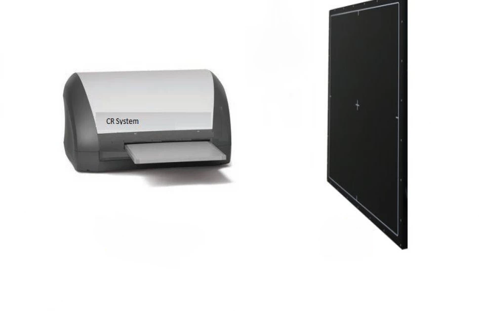
Components of Computed Radiography
Computed Radiography systems comprise several essential elements:
- X-ray Source
The radiation source used for exposing the subject or object to X-rays.
- Phosphor Plates
These imaging receptors replace traditional X-ray film and store X-ray energy using photostimulable phosphor materials.
- Cassettes
Light-tight cassettes enclose the phosphor plates, safeguarding them from light and contaminants.
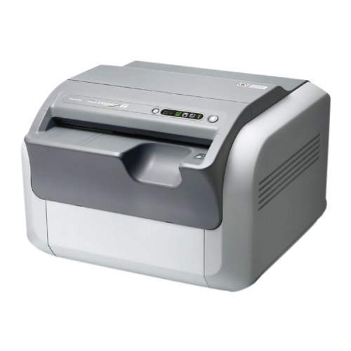
- CR Reader
After exposure, the cassettes are inserted into a Computed Radiography reader. This device includes a laser system for scanning the phosphor plates and a detector for capturing the emitted light.
- Computer System
A computer system processes and displays radiographic images, featuring software for image manipulation, enhancement, storage, and retrieval.
- Display Monitor
Digital radiographic images are presented on a computer monitor for interpretation by radiologists, clinicians, or inspectors.
- Network Integration
Many CR systems are seamlessly integrated into healthcare or industrial networks, allowing effortless image transmission to various destinations, including picture archiving and communication systems (PACS) or remote workstations.
- Quality Control Features
Computed Radiography systems may incorporate quality control features like automatic exposure control and calibration to ensure image accuracy and consistency.
Computed Radiography fulfils essential functions in radiography, encompassing image acquisition, enhancement, storage, transmission, analysis, and dose management.
Its core components include X-rays sources, phosphor plates, cassettes, CR readers, computer systems, monitors, network connectivity, and quality control tools, collectively delivering efficient and high-quality digital radiographic imaging.
Basic Principles of Digital Radiography
Digital Radiography (DR) is a state-of-the-art radiographic imaging technique that has revolutionized the field of medical diagnostics and industrial Non-destructive Testing.
Unlike traditional film-based radiography, Digital Radiography captures and processes X-ray images in a digital format, offering several advantages, including real-time image acquisition, enhanced image quality, efficient workflow, and precise dose control.
DR systems utilize advanced solid-state detectors to directly convert X-ray photons into electrical signals, leading to rapid image acquisition and immediate results.
In this section, we will explore the fundamental principles and components of Digital Radiography, shedding light on its technological intricacies and key functions in the realm of radiological imaging.
Digital Radiography relies on advanced technology to capture and process X-ray images. Its basic principles include:
- Direct Conversion
DR uses solid-state detectors (e.g., amorphous selenium or silicon) that directly convert X-ray photons into electrical charges. This direct conversion process provides high image quality and efficiency.
- Digital Signal
The electrical charges generated by X-ray interaction are converted into a digital signal, creating a digital image with a wide dynamic range and excellent contrast.
- Immediate Image Acquisition
DR systems provide real-time or near-real-time image acquisition. This rapid image capture minimizes patient exposure to radiation and allows for quick assessment.
- Dose Optimization
DR systems can adjust exposure levels in real time, optimizing radiation dose while maintaining image quality.
Function of Digital Radiography (DR) in Radiography
Digital Radiography serves several crucial functions in radiography:
- High-Quality Imaging
Digital Radiography technology offers exceptional image quality, with high resolution and contrast, enabling accurate diagnosis and visualization of anatomical structures.
- Efficiency
Digital Radiography systems provide rapid image acquisition and immediate results, streamlining workflow in healthcare and industry.
- Dose Reduction
Digital Radiography allows for precise dose control, minimizing radiation exposure to patients and workers.
- Image Storage and Retrieval
Digital images are stored electronically, making it easy to archive and retrieve past radiographs for comparison and analysis.
- Digital Enhancement
DR systems offer image processing tools for adjusting contrast, brightness, and other parameters to improve diagnostic accuracy.
Components of Digital Radiography
The key components of Digital Radiography consist of the following:
- X-ray Machine
The X-ray source generates ionizing radiation used for imaging.
- Digital Detector Panel
This panel replaces traditional film or phosphor plates. It directly converts X-ray photons into electrical signals.
- Control Console
The console controls exposure parameters, image processing, and image display.
- Computer System
A computer processes, stores, and displays digital images. It includes software for image manipulation and analysis.
- Monitor
Digital radiographic images are viewed and interpreted on a computer monitor.
- Network Connectivity
DR systems are often integrated into healthcare or industrial networks for image sharing, storage, and transmission.
- Quality Control Tools
DR systems may include features like automatic exposure control and calibration to ensure image quality and consistency.
Digital Radiography relies on the direct conversion of X-ray photons into electrical signals, offering rapid image acquisition, high image quality, and dose optimization.
Its components include the X-ray machine, digital detector panel, control console, computer system, monitor, network connectivity, and quality control tools, collectively providing efficient and high-quality digital radiographic imaging.
Summarization of the Differences between Computed Radiography and Digital Radiography
Computed Radiography
Digital Radiography
Image Capture and Conversion
Uses photostimulable phosphor plates to capture X-ray images and convert them into digital format through laser scanning
Utilizes solid-state detectors (e.g., amorphous selenium or silicon) to directly convert X-ray photons into digital signals, without the need for intermediate plates.
Image Quality
Image quality can be affected by the performance of the phosphor plates and laser scanning, leading to potential variations.
Offers consistent and high-quality images with excellent resolution, contrast, and a wider dynamic range due to direct conversion.
Image Acquisition Time
Slower compared to DR, as it involves the manual handling of phosphor plates and subsequent scanning.
Provides real-time or near-real-time image acquisition, making it faster and more efficient.
Workflow Efficiency
Requires additional steps such as plate handling and scanning, which can introduce delays in the workflow.
Streamlines workflow by providing immediate image results, reducing patient or worker waiting times.
Dose Optimization
Dose adjustment is possible during image processing but may not be as precise as in DR.
Offers real-time dose control, allowing for precise optimization while maintaining image quality.
Maintenance and Durability
Requires maintenance of the reader and the replacement of phosphor plates over time.
Generally, more durable and has fewer components to maintain, reducing long-term costs.
Image Storage and Retrieval
Stores images digitally but may have limitations in terms of accessibility and speed.
Offers efficient and easy image storage, retrieval, and sharing due to its digital nature.
Initial Cost
Typically has a lower upfront cost compared to DR systems.
Initial investment is generally higher but may provide long-term cost savings due to efficiency and durability.
Radiation Exposure
Patient or worker radiation exposure may be slightly higher due to the manual handling of plates and scanning delays.
Allows for precise dose control, minimizing radiation exposure for both patients and workers.
Conclusion
The disparities between Computed Radiography and Digital Radiography manifest in various aspects.
Digital Radiography (DR) notably excels in image quality, acquisition speed, workflow efficiency, and dose optimization due to its direct conversion of X-ray photons into digital signals.
However, it is imperative to emphasize that the choice between Computed Radiography (CR) and Digital Radiography (DR) should not be based solely on these differences.
Factors like budget constraints and specific application needs must be considered when making a decision.
Looking ahead, both Computed Radiography (CR) and Digital Radiography (DR) will continue to hold significant roles in radiography.
Computed Radiography may remain a cost-effective choice in certain situations, while DR is poised for further advancement, potentially integrating innovations such as artificial intelligence for image analysis and optimization.
The future holds promise for the ongoing evolution and enhancement of these technologies, solidifying their vital roles in the field of radiological imaging.
As technology evolves, Computed Radiography and Digital Radiography are expected to synergize and complement each other, offering a diverse toolkit to meet the ever-expanding demands of medical diagnostics and industrial applications.
Their continued coexistence ensures a comprehensive approach to radiographic imaging, addressing a wide spectrum of requirements and challenges in the years to come.
Key Takeaways
Digital Radiography (DR) offers superior image quality, speed, workflow efficiency, and dose optimization due to its direct conversion of X-ray photons into digital signals.
The choice between Computed Radiography (CR) and Digital Radiography (DR) should consider factors such as budget constraints and specific application needs.
Both technologies will continue to play vital roles in radiography, with DR poised for further advancements.
References
1. I.pinimg
2. Hospital store


