

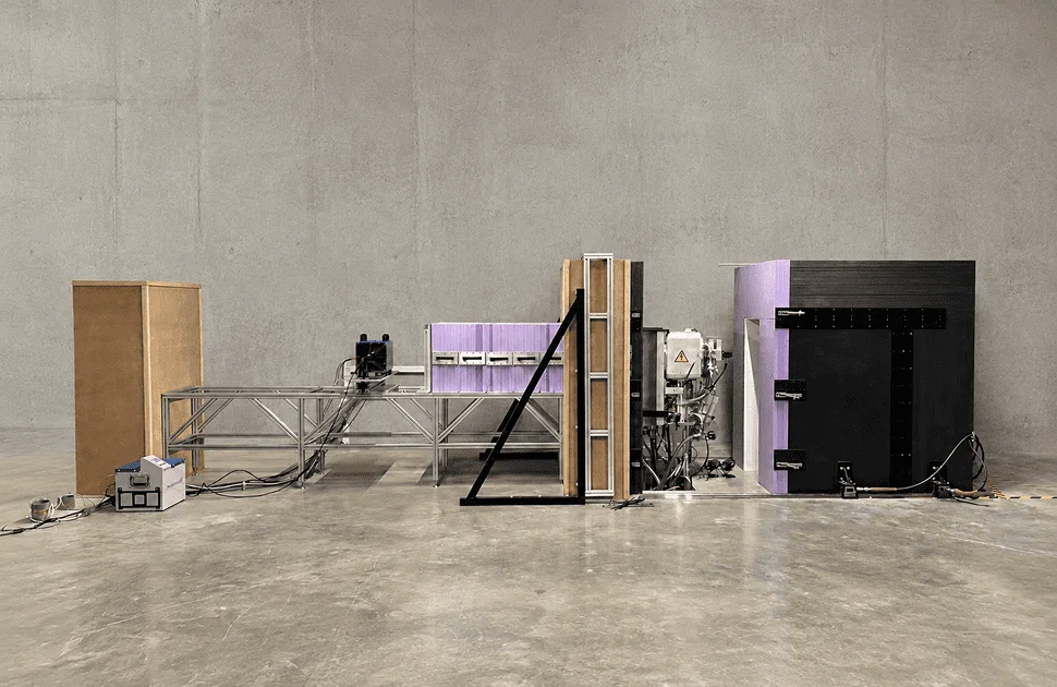
Neutron radiography can reveal materials and features that are hard to see with X-rays. The principle has been known for decades. The challenge has been to bring a useful neutron beam to where industrial inspection takes place. New source, detector and digital imaging technologies are starting to change that.
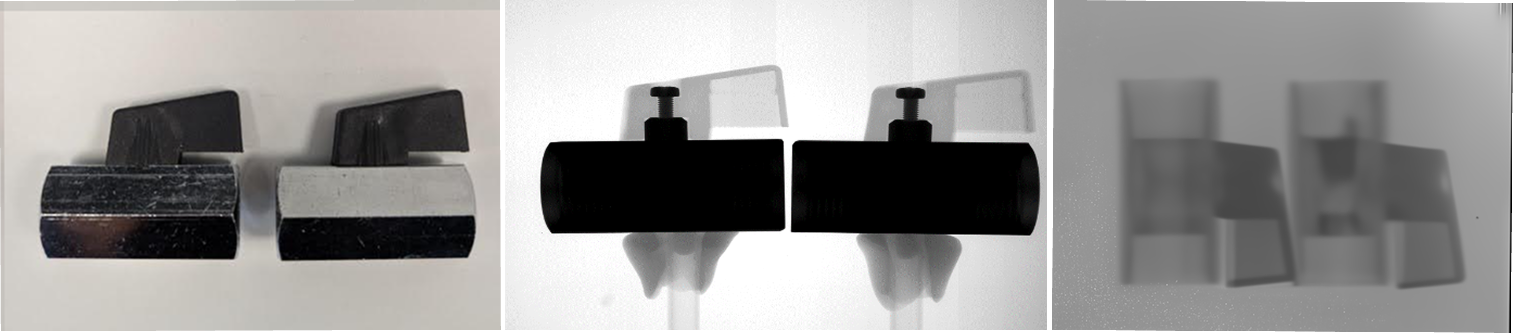
Figure 1. The same two valve parts in a photograph, an X-ray radiograph and a neutron radiograph. The lubricated part is on the right. X-rays map the metal hardware; neutrons reveal the hydrogen-rich grease. Images: Exosens
The quickest way to explain neutron radiography to someone who knows X-ray inspection is to show both images of the same part.
Figure 1 shows two small metal valves, first as a photograph and then in X-ray and neutron radiographs. The valve on the right contains grease; the other does not. In the X-ray image, the metal bodies dominate, and the grease adds little contrast. In the neutron image, the balance shifts. The metal is easier to see through, while the hydrogen-rich grease stands out.
The inspection geometry is familiar. A source illuminates the part, a detector sits behind it, and the transmitted radiation forms a projection image. What changes is the interaction with matter. The two radiographs answer different questions about the same part.
That is the case for neutron radiography in NDT. It does not replace X-ray imaging. It adds another contrast mechanism, and with it another view.
Why the contrast shifts
X-rays interact mainly with the electrons around an atom, so their attenuation tends to rise with atomic number and density. This makes them very good at showing dense metal structures, while many light organic materials are fairly transparent.
Neutrons interact with atomic nuclei instead. Their attenuation follows a much less tidy pattern. Hydrogen interacts strongly with thermal neutrons. Lead and bismuth, both strong X-ray absorbers, are quite transparent to them. Even isotopes of the same element can behave very differently in a neutron beam, while ordinary X-ray radiography offers almost no isotope contrast.
The effect is shown in the scatterplot in Figure 2. If the two modalities attenuated materials in much the same way, the points would gather near the diagonal. They do not. Many sit high on one axis and low on the other.
Neutrons are not just giving us more or less contrast than X-rays. They are giving us different contrast.
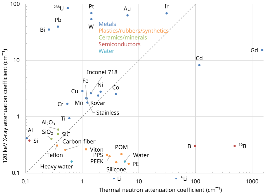
Figure 2. Contrast for thermal neutrons versus contrast for 120 keV X-rays. Materials far from the diagonal differ most strongly in their relative attenuation. Gadolinium is an extreme thermal-neutron absorber. Graphic: Exosens.
“Neutrons are not just giving us more or less contrast than X rays. They are giving us different contrast.”
Gadolinium is the most striking case. It absorbs thermal neutrons so strongly that even a small amount can act as a tag. A gadolinium-bearing penetrant can enter porous material or a fine, surface-connected crack that runs deep into a part. Where the penetrant is retained, the tag leaves a strong mark in the neutron image, even through dense surrounding metal.
The textbook example is ceramic core residue in a high-pressure turbine blade. The porous residue holds the tag and becomes easy to spot. Figure 2 gives the scale of the effect. No element offers X-rays anything close to that contrast on the plotted scale: gadolinium’s thermal neutron attenuation is more than an order of magnitude above the highest X-ray attenuation.
This leads to a useful rule. If an inspection can be done well with X-rays, use X-rays. The method is mature, fast and widely available. Neutron imaging becomes useful when the information sought is a poor match for X-ray contrast. It belongs beside X-ray radiography in the toolbox, not in its place.
The bottleneck is access to sources
Neutron imaging is not new. Modern research facilities can produce neutron radiographs and tomographic data of superb quality. What has kept the method out of day-to-day industrial NDT is the source.
Bright thermal-neutron beams have mainly come from research reactors or large accelerator sites. There are few of them. Beam time must be booked. Parts must be packed and shipped. Lead time, cost, customs, and confidentiality can all become part of what should have been an inspection task.
Established neutron-radiography service providers still record the final image on film. A customer sends in a part; after exposure, the part comes back with physical films. Film can yield excellent images and remains qualified in long-standing processes. Even so, the workflow would be familiar to an X-ray inspector from the middle of the last century.
X-ray inspection has followed a different path. A manufacturer can place a system near production or development and use it when needed. A neutron radiograph has more often meant a special campaign at a remote site.
For Exosens, that led to a different design question. We did not set out to build a small research reactor. We asked how small a neutron source could be made useful for imaging.
Bringing the source into the lab
Neutronis grew from that question. It pairs a compact deuterium-deuterium fusion source with efficient neutron detection and a native-digital imaging chain.
Deuterons are accelerated into a deuterium-bearing target, producing fast neutrons at about 2.5 MeV. A moderator slows them down to thermal energies. Beam-forming and shielding hardware then turn part of the output into the thermal neutron beam used for radiography.
The source can fit into an industrial system. The trade-off is flux: a reactor beamline can deliver orders of magnitude more thermal-neutron flux at the sample. At a bright beamline, even a modest detector can gather enough neutrons for a fine image. With a compact source, every neutron counts. Detection efficiency, beam geometry, shielding and background rejection all carry more weight.
“With a compact source, every neutron counts.”
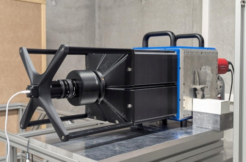
Exosens already had one key part: Neutronic[i], a detector first developed for neutron research facilities. Its high efficiency is a strong match for the lower flux of a compact source. Its camera readout also supports a native-digital chain.
The development prototype pictured at the start of this article brought the source, moderator, beam optics, shielding, detector, and acquisition system together. In an early test, water flowing through channels in an aluminium cooling block became the main feature. The aluminium was fairly transparent. An X-ray image gives almost the reverse balance. The complete chain worked; the next task was to make it useful and repeatable.
Neutronic[i]: making scarce neutrons useful
Most neutron imaging detectors let the great majority of incoming neutrons go unused. Neutronic[i] does not. Its patented neutron-sensitive microchannel plate detects more than half of the thermal neutrons that reach it. That level of efficiency sets it apart from every other neutron imaging detector on the market. At a research reactor, an intense beam can mask detector inefficiency. A compact source has no such luxury. Each neutron captured shortens the path to a clear image; each one missed adds exposure time. Neutronic[i] is therefore much more than the camera at the end of Neutronis. It is the technology that turns the modest flux of a compact source into practical, native-digital neutron radiography on site. TECHNOLOGY BOX |
|---|
When every neutron becomes data
Film is not only a physical medium; it sets the whole service workflow. Neutronis starts native digital. The detector records individual neutron events, and Neutronic[i]’s efficiency allows a high-quality radiograph to be made with much less total neutron fluence than film needs.
That changes what happens after exposure. The image can be corrected, reviewed and shared as data, without developing or shipping physical sheets. Open-beam and background measurements correct for non-uniform illumination and unwanted signal. Filtering, reconstruction and denoising can then work directly on the neutron data. We have also added AI-assisted processing to this set of tools.
Its task is not to invent details that were never measured. Physics still sets how many neutrons reach the detector, and software cannot fix poor statistics. It can, however, make sound use of every event that was recorded. At ASNT in 2025 we showed event-counting acquisition, flat-field correction, advanced post-processing and real-time reconstruction. By ECNDT in 2026, compact-source imaging and AI-assisted denoising were parts of one joined-up chain. A Neutronis image is made by the source, moderator, beam optics, detector, acquisition, calibration and reconstruction working together.
Finding the right inspection problems
A new imaging tool creates a strong urge to point it at everything. We have tried to resist that. The useful question is not, “Can we make a neutron image of this?” It is, “Does this part contain information that another NDT method struggles to show?”
Hydrogen gives some of the clearest examples. We have imaged metal-hydride storage cartridges and could tell whether they were empty, half-full or full through the outer casing. More recently, we imaged a hydrogen fuel cell in operation. Neutrons are a natural fit because they respond strongly to hydrogen and water, both of which can be hard to track with conventional radiography.
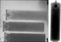
Figure 3. Empty, half-full and full metal-hydride cartridges in neutron transmission, with the X-ray view at right. Images: Exosens.
The same logic reaches into routine mechanical parts. Lubricant inside a heavy assembly may be all but invisible to X-rays yet show strong neutron contrast. Sealants, O-rings, adhesives and polymers can behave in much the same way. Energetic materials are another established field: propellants and explosives contain light elements but often sit inside metal housings. Lithium also makes neutron imaging useful in battery research.
There is no single “neutron application”. These cases share a gap between the information sought and the contrast offered by established methods. That is where another tool earns its place.
From an imaging experiment to an NDT system
There is a wide gap between making a convincing radiograph and delivering industrial NDT equipment. A working system must be safe and predictable outside a neutron facility. Its geometry must be stable and repeatable. Field of view and scan time must fit real parts. Components must be maintained and serviced. Installation and regulatory compliance cannot rely forever on the physicists who built the prototype.
Those tasks now make up much of the Neutronis programme. The generator is being upgraded for more output, the useful imaging area is growing, and the reconstruction chain is improving. In parallel, we are working through the regulatory and system-engineering steps needed for the first customer sites.
There will be trade-offs. A compact source cannot match a major reactor, and some work will still need a research beamline. But a reactor beamline in a smaller box was never the goal. The goal is enough image performance in a system that can sit where the inspection problem is.
“A reactor beamline in a smaller box was never the goal.”
A second view of the same part
An NDT engineer might use X-rays to map the metal in a sealed assembly, then use neutrons to show where a fluid or seal sits inside it. A developer working on a hydrogen system might use one modality for the hardware and the other for hydrogen or water. Neither image is better. Each answers a different question.
For decades, that second image has meant sending a part to one of a few specialist sites. Compact neutron generators, efficient detectors and native-digital imaging now offer a way to change that model.
If we succeed, the most important change may not be any single Neutronis specification. It will be that, when an X-ray image leaves an NDT engineer with an unanswered question, asking for a neutron image no longer sounds exotic. It will simply mean reaching for another tool.
About The Author

Serge Duarte Pinto - Director of Innovation, Exosens
Serge Duarte Pinto develops particle detectors and neutron-imaging systems for scientific and industrial use. He holds a PhD in high-energy physics from the University of Bonn and a Global Executive MBA from Rotterdam School of Management. His earlier work at CERN and TU Delft focused on radiation detectors and neutron instrumentation. He leads the Neutronis programme. More: exosens.com/neutronistm
There are no comments yet!