
Table Of Contents
- Introduction to Radiographic Testing and Image Quality
- What Are Penetrameters and IQIs?
- Types of IQIs and Penetrameters
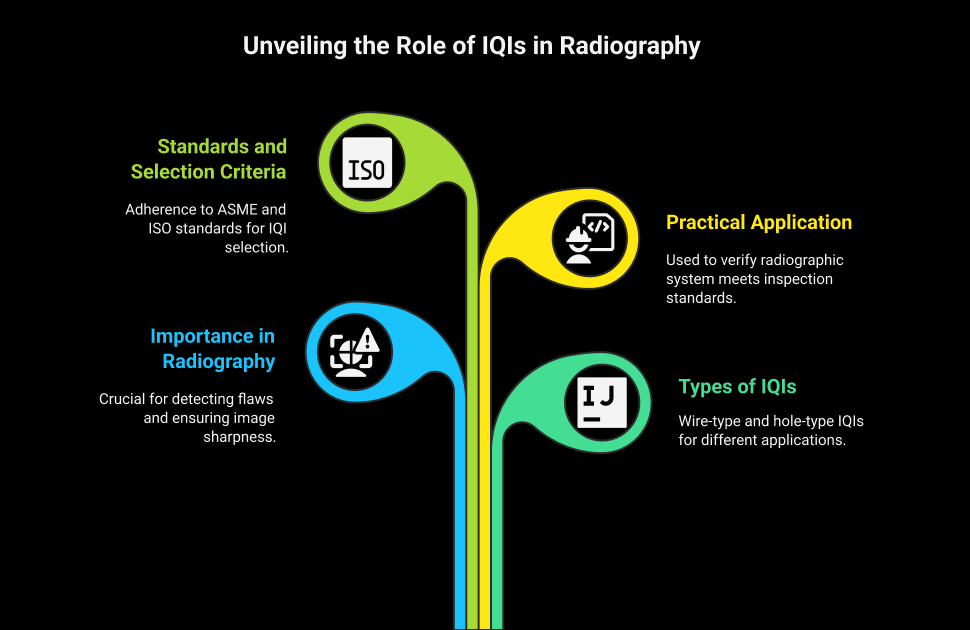
- Standards and Selection Criteria
- Practical Application in Radiographic Testing
- Radiographic Techniques and IQI Integration
- Evaluating Radiographs with IQIs
- Advanced Considerations
- Conclusion
- FAQs
Introduction to Radiographic Testing and Image Quality
In a construction site, inside one of the departments of a massive expansion project of a refinery, a radiographic technician places a very thin metal strip with very fine wires printed on it over a newly welded pipeline. Apparently, it is not so simple; after all, it is merely a label, a ritual. Superficially small strips, called a radiographic penetrameter, will decide whether a radiograph made in less than ten minutes will have sufficient credibility, or there may be a deadly flaw inside which will go unnoticed. Behind every successful radiographic testing procedure lies a quiet but powerful standard: the Image Quality Indicator (IQI), a cornerstone of image quality indicator radiography.
In radiographic testing (RT), where X-rays or gamma rays are used to detect internal discontinuities like voids, inclusions, or cracks in welds and materials, image quality is everything. Observing a flaw is not only about having the appropriate exposure, it is about the knowledge that the picture is sharp enough to display the most minor of dangers. This is where IQIs - also known as penetrameters- can be used. These architectured precision tools do not detect imperfections, but they make sure your imaging system can. They simulate flaws of known dimensions, helping radiographers verify that the system meets the minimum IQI standards ASME or ISO 19232 requires. Understanding penetrameter use in RT is crucial for confirming whether the radiographic procedure truly meets inspection standards.
The real essence of the IQIs and penetrameters is that they offer a quantification of the image quality in a quantitative manner. In their absence the proper radiographic technique would give erroneous results at the best and, particularly when it would be a planar defect or a complicated weld shape that was involved, or when use was to be made of a dense material, knowledge of the location and quantity of the defect would be of the essence in the matter of radiography. For NDT professionals and inspectors, understanding how to select, position, and interpret IQIs—and the different IQI types RT offers—is as important as knowing how to operate the radiography equipment itself. This knowledge forms the foundation of any trusted radiographic penetrameter guide used in field or code-based applications.
In the pages ahead, you’ll explore the fundamentals and advanced concepts behind penetrameters and IQIs in radiographic testing. You will get to know what they are, the distinction between the wire-type IQIs and hole-type IQIs, as well as how they are applied in different standards. Selection criteria, placement techniques, and their validation in verification of radiographic sensitivity will also be walked through the article. Is it an inspection, exam preparation, or all-around quality control improvements you have in mind? No matter the case, this guide will provide you with the clarity you need to apply IQIs appropriately and accurately such that your radiographs are well within industry expectations and, much more importantly, pick up what counts.
What Are Penetrameters and IQIs?
The European name used to describe penetrameters is Image Quality Indicator (IQI). It is a precision engineered instrument that is attached to a test object during radiographic exposure to assess the ability of the radiograph to reveal a defect. The tools offer an objective measure of image quality which depends on three major factors: unsharpness (blurring), contrast (density change because of change in thickness) and noise (e.g. graining of film). IQIs are not designed to assure that they will identify every flaw in the network, especially the smaller or defectively oriented ones such as cracks, but will provide an assurance that the radiographic process has achieved a baseline quality.
IQIs normally come in materials radiographically comparable to the examined item like steel, aluminum, titanium or copper, to echo radiation consumption proclivities of the specimen. They are made in a way that they are able to simulate defects so that radiographers can quantify the smallest detectable feature as a percentage of the thickness of the material. The sensitivity level attained is represented by the smallest visible part on the radiograph that may be either a wire or hole.
Types of IQIs and Penetrameters
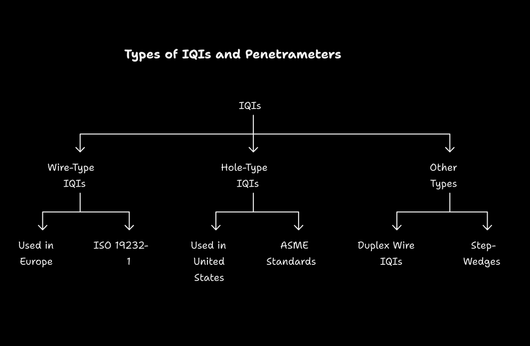
IQIs come in two main forms in the field of industrial radiography namely: wire-type and hole-type (which also come under step-hole or plaque-type). Both types are used in measuring the radiographic sensitivity and each is chosen depending on the relevant standards to be used and the test material.
1. Wire-Type IQIs
The most common form of IQI is a wire-type, which is commonly used in Europe and is standardised under ISO 19232-1 (previously EN 462-1) and is wired together in seven parallel wires of smaller and smaller diameter bound in plastic. The wires can be formed of such materials as steel (Fe), aluminum (Al), titanium (Ti), or copper (Cu) that are identical with those of the test object. Quality can be determined right away by the thinnest visible wire on the radiograph where the determination is quoted as a percentage of the material thickness. Take, e.g., 0.2mm wire on a 10mm thick specimen would mean sensitivity of 2%. Wire-type IQIs are ideal when pipeline inspectors need to check the detectability of linear imperfections such as piping.
2. Hole-Type (Step-Hole) IQIs
Hole-type IQIs (which are sometimes used in the United States, and are known as penetrameters to ASME standards), are thin plates with three holes of diameter of one, two and four times the plate thickness (1T, 2T and 4T holes). Normal thickness of a plate is 2 percent of the thickness of the specimen and the tiniest hole which could be observed represents the level of sensitivity. As an example, a 2-2T quality indicates that the penetrameter is 2 percent of the thickness of the specimen, and the 2T hole (twice the plate thickness), is visible. These IQIs are rectangular and are available in two dimensions of 0.5 inch by 1.5 inch of material up to 2.5 inches thick or 1 inch by 2.25 inch dimensions of material greater than 2.5 inches thick. The holes are a loss of material that creates holes to simulate holes or inclusion.
3. Other Types
Rarer types are duplex wire IQIs (ISO 19232-5) which measure absolute unsharpness and step-wedges which can be used in parts of the world such as Japan or France to measure contrast and radiation energy (kV). The tools are additional solutions to make sure that particular radiographic parameters are achieved.
Standards and Selection Criteria
International standards are used to apply IQIs like ASME Section V, ISO 19232, and EN 462 that describe requirements on IQI material, location and sensitivity. Some of the considerations are:
- Material Selection: The IQI needs to be of the same alloy group or of another material that has a lower radiation absorption capacity in comparison to the test object. This makes it able to represent the radiographic property accurately of the specimen.
- Position: IQIs may be positioned on the source side of the test object so as to create the worst possible situation of detection. When the film side of the film needs to be put, then a lead marker labeled F is placed to signify the deviation.
- Sensitivity Conditions: The greatest use of penetrameters is most industrial applications which dictate a 2-2T sensitivity where the penetrameter thickness will be 2% of the specimen thickness, and the 2T hole or equal wire will be visible. The image quality is better depicted by higher sensitivity (less percentage).
- Standards Compliance: Wires size and holes are also defined by such standards as ASME Section V and ISO 19232-1. Case in point, four types of wire-type IQIs have been defined in ISO 19232:1 and consist of a set of wire combinations that can be used in every inspection.
Practical Application in Radiographic Testing
Practically, IQIs are located on the periphery of the radiation field centerline which is of low intensity position so as to confirm that a defect within the region of interest is detectable. The radiograph with the penetrameter demonstrates that the exposure methods, type of film and the conditions of processing attain the desired sensitivity. As an instance, in a single-wall single-image (SWSI) technique, radiation will be transmitted through one thickness material, and the contrast image will be produced. Smaller components (diameters 1/lee 3.5 inches) can use a double-wall method such as double wall, double-image (DWDI) or double-wall single-image (DWSI) and this will need multiexposure to cover the weld edge.
Another measure is the radiographic density, which determines the darkness of the film. Standard code requirements denote the ranges of density of 1.8-4.0 on X-ray and 2.0-4.0 on gamma ray, which establishes enough visibility so as to be able to detect defects. The density and the contrast are on standard because the outline of the IQI and the holes/wires are visible.
Limitations of IQIs and Penetrameters
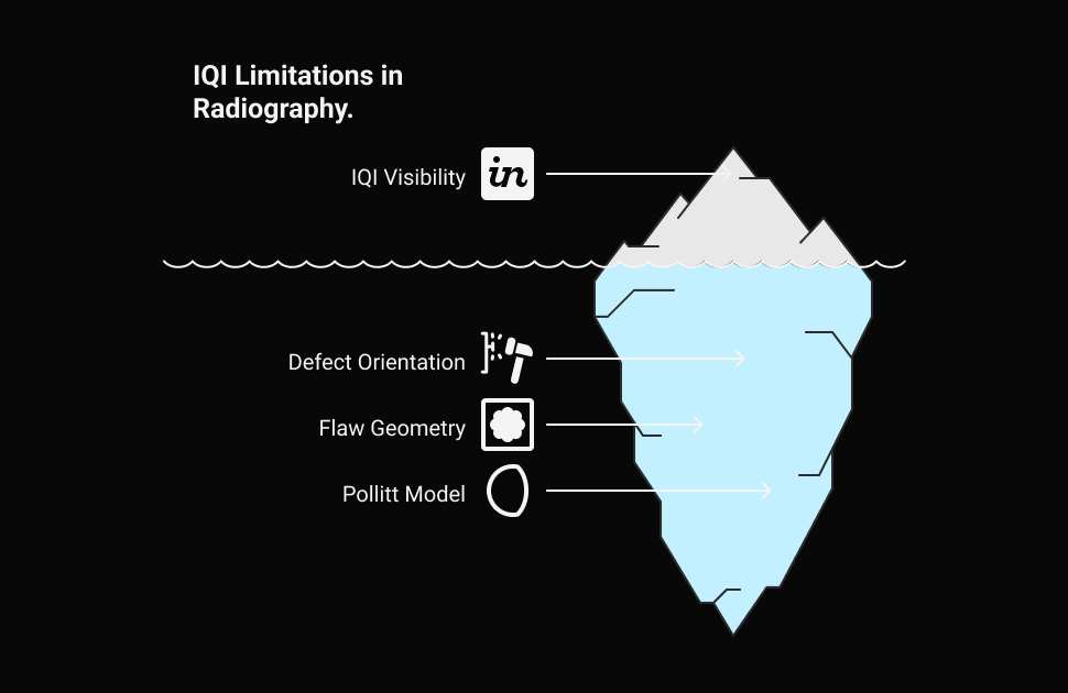
IQIs are necessary when it comes to the validation of radiographic quality but come with their limitations. It is the view of objects that does not assure the view of defects of similar dimensions whether the latter are planar such as cracks or the absence of fusion. The detection of these defects is dependent on their orientation to the beam of the radiations as a vertically oriented flaw is more difficult to detect as there is hardly any change in density. The Pollitt model points at the sensitivity of the flaws as something that is affected by the aspects such as the fault gap and beam angle, which are not replicated fully by the IQIs. Moreover, the actual defects do not possess regular geometrical shapes such as those of the standardized holes or wires on the IQIs, and it is difficult to have direct correlation thereon.
Radiographic Techniques and IQI Integration
The radiographic technique has a considerable bearing on the IQI effectiveness. SWSI technique is favored because of its simplicity and clarity but complex geometry may be treated using a double-wall technique because of pipes. In DWDI, the rays are radiated through both walls thus creating an elliptical image whereas DWSI radiates on one thickness of weld. Radiograph also contains location markers and identification info made of lead which is to maintain traceability without obstructing the region of interest.
When needing a source with a certain sensitivity the required radiation source is chosen depending on the thickness of the material to be tested (X-rays or gamma e.g. Iridium-192 between 6.3--76.2 mm thick steel or Cobalt-60 between 38 and 178 mm). The IQI makes sure that the obtained source and technique will produce the appropriate image quality.
Evaluating Radiographs with IQIs
The last stage of RT is conducting an expert examination of the radiograph. The interpreter evaluates the IQI visibility to find out the radiograph complies with the necessary sensitivity (e.g., 2-2T in ASME standards). They also take into account welding processes and related discontinuities including lack of fusion in arc welding, which might be not easily detected when it is in an unfavorable direction. The IQI serves to give a numerical value of what constitutes the status of image quality but the skill of the interpreter is paramount in discovering the real defects.
Advanced Considerations
G/σ_D (gradient-to-noise ratio) is an objective parameter to distinguish among the film processing systems and assure the dissimilar image quality along with the variances related to the film density. In the case of digital radiography, the sensitivity of IQIs is of concern, but the other aspects, which are reviewed, are the resolution of the detector and the signal-to-noise ratio. Such standards as BS EN ISO 19232-3 outline sensitivity requirements including a 0.8 mm step/hole equivalence for 25 mm thick materials when using SWSI techniques with adaptation to thinner materials or DWDI apparatus.
Conclusion
Penetrameters and IQIs are indispensable tools in radiographic testing, providing a standardized method to assess image quality and ensure reliable defect detection. They quantify the radiographic sensitivity by simulating flaws by using wires or holes, and one generally seeks to achieve a 2% thickness detectability. They do not ensure that all the defects can be identified but when used well using standards such as ASME and ISO then they improve the confidence in the RT inspection. For NDT professionals, researchers, and students, understanding the selection, placement, and limitations of IQIs is crucial for mastering radiographic testing and ensuring the integrity of critical components.
FAQs:
1. What is the use of a penetrameter in radiography?
It is a tool used to measure the contrast or density change in an image given a known change in specimen thickness. The penetrameter is a useful way to assess the overall caliber of the radiography examination.
2. What is the IQI in radiography testing?
The IQI is not meant to be used as a gauge of the size of a cavity that may be visible on the radiograph; rather, it is used to show the quality of the radiography technique.
3. What is the purpose of IQI?
Sensitivity Values of the IQI: Assess the sensitivity of a radiography technique using Image Quality Indicators (IQIs), which are sensitivity levels reported as a percentage of the object's thickness.
4. How to calculate IQI?
The thickness of the radiographed plate divided by the diameter of the smallest IQI wire visible, x 100. Therefore, from a sensitivity perspective, the radiography film is appropriate because the sensitivity is more than what is needed.
5. How do you select IQI in radiography?
The thickness of the component and the type of metal are taken into consideration when choosing the IQI; the thicker the component, the thicker the IQI's wires.


