
Table Of Contents
- Introduction
- Evolution of Digital Radiographic Image Interpretation
- Human Limitations in Radiographic Interpretation
- AI’s Role in Enhancing Digital Radiographic Interpretation
- Image Processing and Feature Extraction in AI-Driven Interpretation
- AI Algorithms for Defect Detection
- Challenges in Implementing AI in Radiographic Interpretation
- Practical Applications in Digital Radiographic Interpretation
- Future Trends in AI-Driven NDT
- Conclusion
- FAQs
Introduction
Digital radiography (DR) is an advanced form of radiographic testing (RT) that uses digital detectors instead of conventional film to capture high-resolution X-ray or gamma-ray images for non-destructive testing. These systems offer real-time image capture, processing and evaluation, with better contrast, wider dynamic range and more efficient storage than film. The interpretation of those images, however, still depends on human judgment — and is therefore exposed to visual fatigue, cognitive bias and subjectivity. Artificial intelligence addresses those limitations directly, applying machine learning, deep learning and computer vision to automate defect detection, improve consistency and shorten evaluation time.
AI-assisted radiographic interpretation uses convolutional neural networks (CNNs) and pattern recognition to identify discontinuities such as cracks, porosity, inclusions and lack of fusion — including indications that are difficult to resolve by eye. These models are trained on large volumes of annotated radiographs, which improves their ability to separate genuine discontinuities from artifacts while keeping evaluation consistent from image to image. As industry moves toward NDT 4.0 and predictive maintenance, AI in radiography supports not only reliability and safety but also data-driven decisions and real-time inspection analysis.
This article covers how film-based interpretation evolved into AI-assisted digital systems, where human interpretation breaks down, how AI addresses those gaps, which algorithms process the images and classify the indications, the practical applications across industry, the obstacles to adoption, and where the technology is heading.
Evolution of Digital Radiographic Image Interpretation
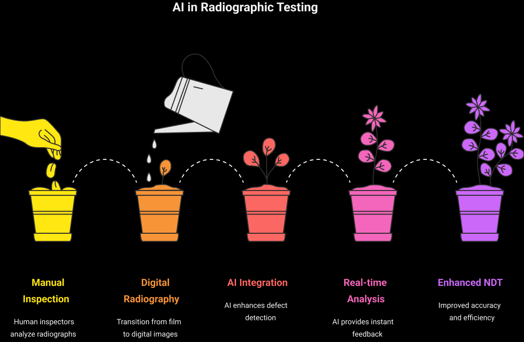
Radiographic interpretation in NDT was performed on analog film for most of the twentieth century. Film review was slow, dependent on the individual interpreter, and impossible to automate. The arrival of computed radiography (CR) in the early 1980s, followed by digital detector arrays (DDAs) over the following two decades, changed that: once the image existed as data, it could be processed, enhanced and analyzed by software.
That shift created the basis for automated defect recognition (ADR). First-generation ADR systems in the 1990s were rule-based, applying classical image processing to detect features such as porosity or cracks in high-volume applications like automotive castings. They improved throughput, but they were rigid — each new part, material or exposure condition required manual re-tuning of thresholds.
Machine learning and deep learning changed the economics of that problem. Rather than encoding rules, AI-driven ADR learns from data and adapts to variation in defect appearance and imaging conditions. CNNs in particular are effective at recognizing complex patterns in X-ray images and at segmenting indications from background. In safety-critical sectors such as aerospace and oil and gas, this allows AI to take over routine screening while the certified interpreter concentrates on evaluation and disposition.
The standards framework has kept pace with the imaging technology, if not yet with the AI layer on top of it. Digital radiographic practice is covered by documents including ASTM E2033 for computed radiography, ASTM E2698 for examination using digital detector arrays, ASTM E2737 for DDA performance evaluation and long-term stability, and ISO 17636-2 for radiographic testing of welds with digital detectors.
Human Limitations in Radiographic Interpretation
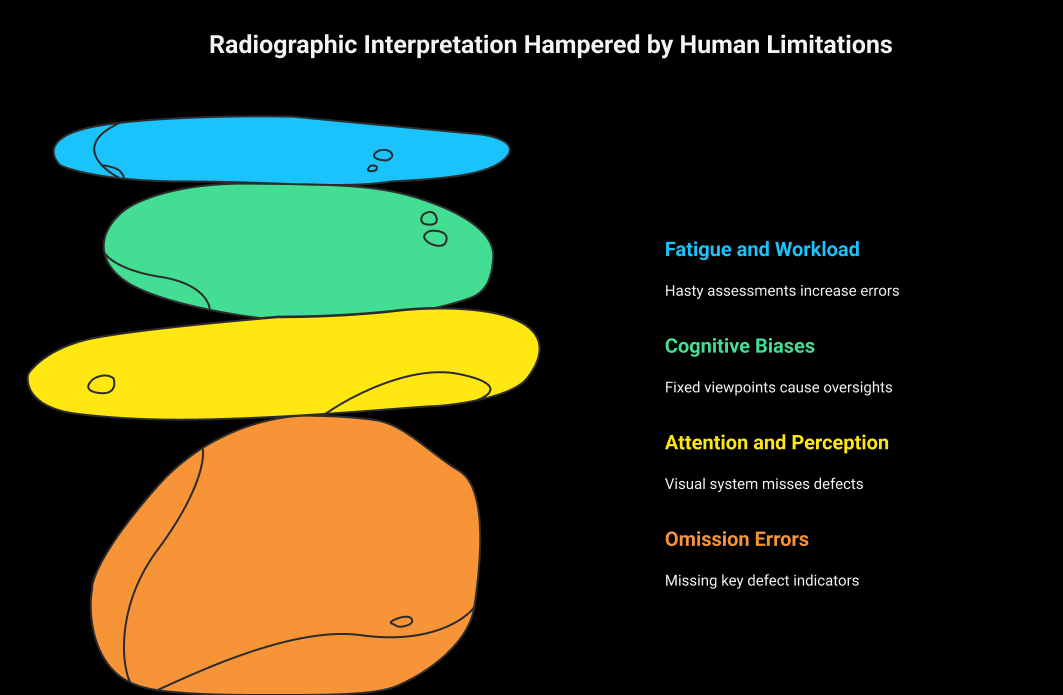
Interpreting radiographic images is a specialized skill that requires trained, qualified personnel. Even experienced interpreters face constraints that affect reliability:
- Omission errors: An interpreter may fail to search a region thoroughly, fail to recognize an indication as significant, or misclassify a genuine indication as an artifact. These are commonly described as search, recognition and decision errors.
- Attention and perception: The visual system samples only part of an image at a time. An indication that is obvious once pointed out can go unseen when it falls outside the search pattern.
- Cognitive bias: Anchoring bias fixes the interpreter on an initial assessment. Availability bias draws attention toward recently encountered defect types, which can leave rarer discontinuities under-weighted.
- Fatigue and workload: Long review sessions degrade accuracy, and both false calls and missed indications tend to rise. Heavy caseloads, tight schedules and interruptions compound the effect.
None of this makes human interpretation unreliable as such — it is the basis of every qualified RT program. It does mean that performance varies with conditions in ways an automated system does not, which is where AI adds value as a support to the interpreter rather than a replacement.
AI’s Role in Enhancing Digital Radiographic Interpretation
AI supports digital radiographic interpretation in four main ways:
- Detection support: Deep learning models, particularly CNNs, can flag small or low-contrast indications such as micro-cracks, fine porosity and inclusions that are demanding to resolve under time pressure. Published results in this area are promising, but performance is specific to the material, defect type and imaging setup on which the model was trained.
- Speed and throughput: Automated pre-screening processes large image volumes far faster than manual review, which matters in high-output environments such as automotive manufacturing. Faster turnaround also makes it feasible to feed results back into process parameters while a production run is still going.
- Consistency: An AI model applies the same criteria to every image. Human evaluation varies across shifts, operators and workload, so automated pre-screening narrows that spread and gives less experienced interpreters a consistent reference.
- Classification and process insight: Beyond detection, models can classify indication types — lack of fusion, slag inclusion, porosity — and aggregate the results. Trends in weld defect type and location can then point back to the welding parameters producing them.
Used this way, AI reduces rework and scrap by catching process drift earlier. It also combines with computed tomography (CT) to add three-dimensional context, and supports predictive maintenance by tracking how indications develop over successive inspections.
Image Processing and Feature Extraction in AI-Driven Interpretation
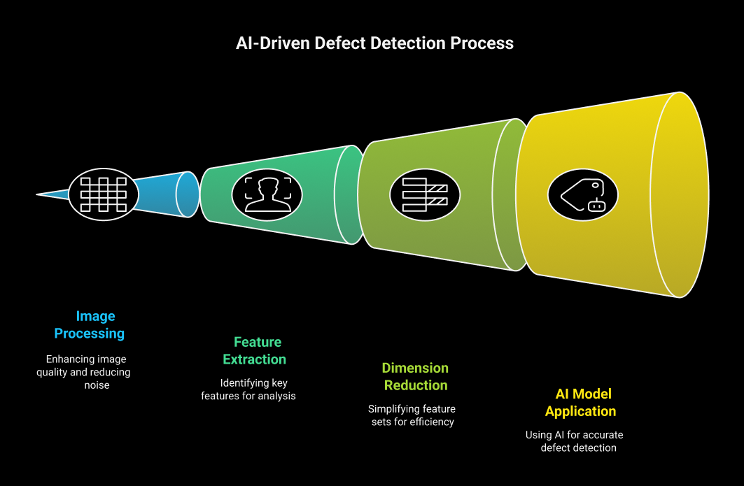
Industrial radiographs frequently suffer from low contrast, noise and uneven grayscale distribution, all of which can obscure indications. Image processing and feature extraction prepare the image so that defects become separable from the background.
- Image processing techniques: Histogram equalization redistributes pixel values to improve contrast. Median filtering suppresses noise while preserving edges. Wavelet transforms decompose the image into frequency components so noise can be removed selectively. Adaptive thresholding separates indications from a varying background, and morphological operations clean up defect geometry — for example, closing gaps within a cluster of porosity.
- Feature extraction: Intensity-based features such as mean and standard deviation describe grayscale variation. Texture features such as entropy quantify local randomness, which helps distinguish genuine discontinuities from artifacts. Edge-based features derived from Sobel or Canny operators pick out crack-like geometry, and shape descriptors such as area, perimeter and aspect ratio support classification.
- Dimensionality reduction: Methods such as principal component analysis (PCA) reduce the size of the feature set, cutting computational load while retaining the information that carries discriminative value.
These processed features feed the classification stage. CNNs compress this pipeline by learning feature extraction directly within their convolutional layers, working from the raw image rather than from hand-engineered descriptors — an advantage in complex weld inspection where defect appearance varies widely.
AI Algorithms for Defect Detection
Several classes of algorithm are used in automated radiographic interpretation, and the right choice depends on the data available and the decision required.
- Classical machine learning: Support vector machines (SVMs) suit binary separation — defect against non-defect — on reasonably balanced datasets. K-nearest neighbors (KNN) handles multi-class problems such as distinguishing porosity from inclusions.
- Ensemble methods: Random forests and gradient boosting combine multiple weak models into one stronger predictor. They tend to generalize better on the imbalanced datasets typical of NDT, where defective parts are the minority class.
- Deep learning: CNNs perform the segmentation task, and architectures such as U-Net delineate defect boundaries so that extent can be estimated. Mask R-CNN adds instance-level segmentation, giving pixel-accurate measurement of individual indications. Transfer learning from pre-trained backbones such as ResNet reduces the volume of annotated data needed to reach usable performance.
- Emerging models: Multimodal large language models are being explored for reporting support — generating narrative descriptions of detected indications to accompany the quantitative output.
Multi-scale approaches such as feature pyramid networks help where defects vary in size within the same image, and instance segmentation provides the precise localization needed for disposition. Together these methods automate the routine screening work and leave the interpreter to concentrate on evaluation against the acceptance criteria.
Challenges in Implementing AI in Radiographic Interpretation
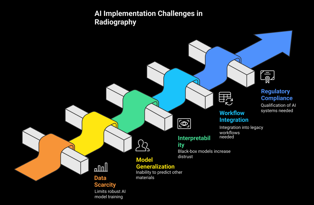
AI in radiographic interpretation faces real constraints, and most of them are practical rather than algorithmic.
- Data scarcity: Industrial defect data is usually proprietary, and the rarest defect types are by definition the least represented. Synthetic data from simulation, such as Monte Carlo X-ray modeling, fills part of the gap, but a domain shift remains between simulated and real radiographs.
- Model generalization: A model trained on aluminum castings will not necessarily transfer to steel welds. Differences in material, thickness, exposure geometry and defect morphology all degrade performance, which makes consistent imaging parameters and preprocessing essential.
- Interpretability: Black-box outputs are difficult to accept in a discipline built on documented, defensible evaluation. Explainable AI methods that show which image regions drove a decision are a prerequisite for wider trust.
- Workflow integration: AI tools have to fit existing inspection workflows, which often involve legacy acquisition hardware and established reporting software.
- Data security: Radiographic data can be commercially or operationally sensitive, particularly in aerospace and defense, so storage, transfer and model-training arrangements need to be controlled.
- Liability: Responsibility for a missed indication does not transfer to a software vendor. This argues for conservative deployment, with AI used as a screening and support tool rather than as the final evaluation.
Qualifying AI within an NDT quality system
The question most often raised by Level IIIs is not whether AI works, but how it is qualified and who remains accountable. A defensible deployment generally has to address:
- Measured performance, not vendor claims. Probability of detection and false call rate should be established on representative parts, materials and defect types, using an accepted methodology such as that set out in MIL-HDBK-1823A.
- Defined role in the workflow. Whether the system pre-screens for a human interpreter, performs a second review after evaluation, or operates as an accept/reject gate changes the evidence required to justify it.
- Retained human accountability. Personnel qualification schemes such as SNT-TC-1A, ANSI/ASNT CP-189, ISO 9712 and NAS 410 place responsibility for interpretation on a certified individual. An AI output does not discharge that responsibility, and the report is still signed by a qualified person.
- Revision control over the model. A retrained model is a changed inspection system. Model version, training set and validation results belong in the equipment and procedure records like any other controlled item.
- Procedure and customer approval. The written procedure has to permit the technique, and customer or code requirements may need addressing before AI-assisted evaluation is accepted on qualified work.
Treating an AI tool the way any other piece of inspection equipment is treated — identified, qualified, documented, revision-controlled — is what converts a promising capability into an auditable one.
Practical Applications in Digital Radiographic Interpretation
AI-assisted digital radiography is in use across several sectors, each with a different driver:
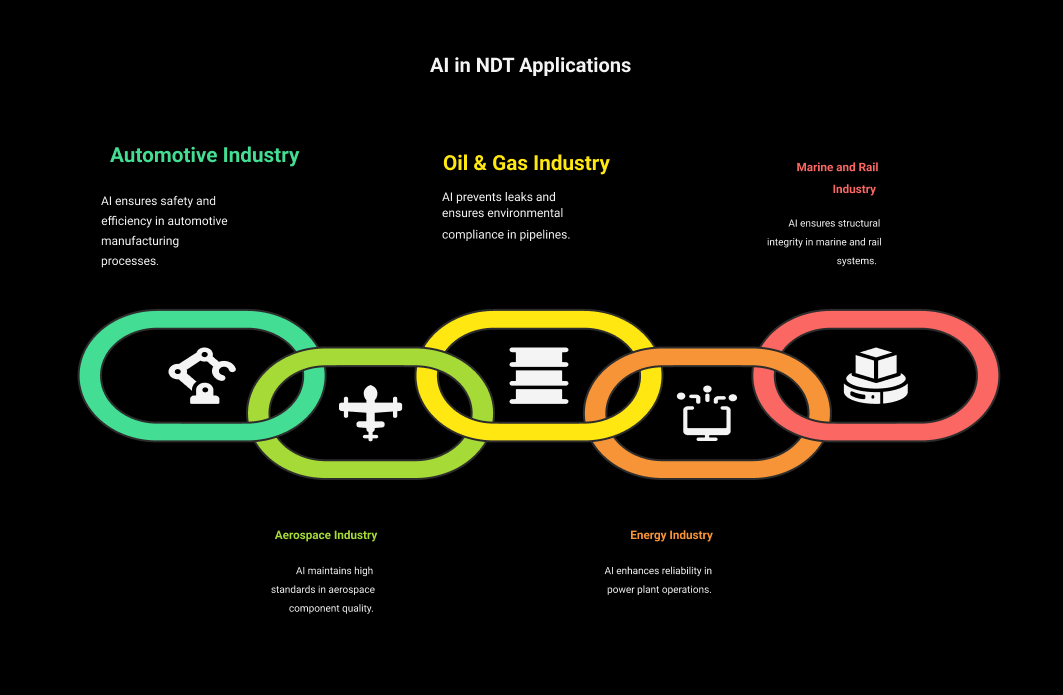
- Automotive: Screening of engine castings and welds for porosity and cracks, reducing scrap on high-volume lines where manual review cannot match production rate.
- Aerospace: Examination of turbine blades, airframe structures and laser welds against demanding acceptance criteria. Combining multiple models reduces uncertainty and helps suppress false calls that would otherwise generate unnecessary rework.
- Oil and gas: Evaluation of pipeline girth welds, supporting integrity management and environmental compliance, with analysis available close to real time in field conditions.
- Power generation: Detection of cracking in boiler tubing and cast turbine components, supporting plant reliability programs.
- Marine and rail: Examination of hull welds and rail components as part of structural integrity assessment.
Cloud deployment allows remote review by specialists who are not on site, and integration with robotic and in-line systems supports process correction during production, consistent with the NDT 4.0 model.
Future Trends in AI-Driven NDT
- Predictive analytics: Tracking indication trends across inspections to anticipate process drift or equipment wear, shifting radiography from detection toward prevention. Weld defect patterns, for example, can signal the need to adjust welding equipment before rejects appear.
- Advanced architectures: Transformer-based models capture longer-range image relationships than convolutional networks, and unsupervised anomaly detection offers a route to flagging unfamiliar defect types without labeled examples.
- Explainable AI: Explanation modules that show the basis for a decision will be central to acceptance, both for interpreter trust and for audit defensibility.
- Data-centric development: Curated datasets, synthetic data generation and domain adaptation are doing more for model robustness than architecture changes. Digital twins can simulate realistic radiographs covering rare conditions that are difficult to capture physically.
- Standardization: Work within bodies such as ASTM and ISO on qualifying automated and AI-assisted evaluation — performance thresholds, validation methodology and human-in-the-loop requirements — will determine how quickly the technology can be used on code work.
- Workforce development: Training in the supervision of automated systems is likely to become part of NDT qualification, preparing interpreters to work alongside these tools rather than around them.
These developments align with Industry 4.0, where inspection data feeds directly into connected manufacturing environments for real-time quality control.
Conclusion
AI is changing how digital radiographic images are interpreted in non-destructive testing. It improves consistency of defect detection, shortens evaluation time and produces data that can be fed back into process improvement. Sectors from automotive to aerospace and oil and gas already use it to assess component integrity, supporting quality and safety requirements while reducing cost and enabling predictive maintenance.
The constraints are equally real: limited training data, generalization across materials, explainability, and the qualification and liability questions that come with using an automated system on code-governed work. Progress on algorithms, synthetic data and standardization is narrowing each of them.
The realistic near-term position is not AI replacing radiographic interpreters but extending what a qualified interpreter can cover reliably. Organizations evaluating these systems can compare digital radiography equipment and ADR software or find NDT service providers and solution vendors working in this area.
FAQs
1. Is radiography a non-destructive testing?
Yes. Radiography uses X-rays or gamma rays to examine a component for internal discontinuities without damaging or altering it, which is what makes it non-destructive. It is one of the volumetric NDT methods, alongside ultrasonic testing.
2. How is AI used in radiography?
Mainly for automated defect recognition. Models trained on annotated radiographs screen incoming images, flag candidate indications such as porosity, cracks, inclusions and lack of fusion, classify them by type and estimate their extent. In most deployments the output is a pre-screen or a second review that a certified interpreter then evaluates against the acceptance criteria.
3. What are the non-interpretive uses of AI in radiology about?
No, not under current qualification schemes. SNT-TC-1A, ANSI/ASNT CP-189, ISO 9712 and NAS 410 place responsibility for interpretation and reporting on a certified individual, and an AI output does not transfer that responsibility. AI is applied as a support tool, with the qualified interpreter retaining evaluation and sign-off.
4. How accurate is AI radiology?
It depends on the material, defect type, imaging setup and the data the model was trained on, so a single accuracy figure is not meaningful. What matters for industrial use is measured probability of detection and false call rate on representative parts, established using an accepted methodology rather than taken from vendor literature. Performance also degrades when a model is applied outside the conditions it was trained on.
5. What are the disadvantages of AI in radiology?
The main ones are limited and proprietary training data, poor transfer between materials and imaging conditions, difficulty explaining how a decision was reached, integration with legacy inspection workflows, and unresolved questions about qualification and liability when an automated system contributes to a code-governed evaluation. Data security is a further consideration in aerospace and defense work.

There are no comments yet!